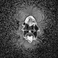
Time-Lapse Revealing Water Patterns Of Starfish Larva Wins Nikon Small World In Motion Competition

Posted on December 14, 2016
Nikon Instruments Inc. today unveiled the winners of the sixth annual Nikon Small World in Motion Photomicrography Competition, awarding First Place to William Gilpin of Stanford University for his video depicting an eight-week-old starfish larva churning the water around its body as it searches for food. Gilpin and his colleagues studied the starfish larva as a model system for how physics shapes evolution, and were surprised and intrigued that a common organism like a starfish could create such an intricate and unexpected pattern in the water.
These complex currents are not only aesthetically pleasing – they depict a phenomenon that was previously unknown, and illustrate how starfish larvae have evolved intricate appendages to create these beautiful and physically taxing flow patterns. The elegant vortices of water efficiently pull particles towards the animal’s body, but it comes at a price: reducing the larva’s ability to swim to escape predators while also broadcasting its current location.
This microscopic larva, although only less than a millimeter in length, could have big implications. The cilia in the starfish larva that make these vortices possible are universal and present in most other organisms, including humans. “While starfish are the among the first animals that have evolved to control the environment around them in this manner, science proves that adaptations are likely mimicked by other more-complex animals later,” said Gilpin. “Biology aside, this process can also be the foundation for industrial purposes for something like advancements in water filters for precise manipulation of water.”
To create the video, Gilpin and his team used dark field microscopy to film the paths of small plastic beads that were directed by the flow currents around the starfish, similar to how photographers capture time-lapse videos of star trails in the night sky. They then stacked images in contiguous groups to make a time-resolved long-exposure video to showcase the movement.
To capture and share movement – and life - seen under the microscope is a relatively new phenomenon, powered by advances in video capabilities in recent years. This is only the sixth year of the “In Motion” category, led by scientists and artists like Gilpin pulling back the curtain on a dimension of life previously unseen to all but a select few.
“The beauty of this time-lapse video and the science behind it epitomizes how video is not only essential to scientific researchers, but to inspire future scientists to explore life around them,” said Eric Flem, Communications Manager, Nikon Instruments. “It is one thing to see a still image captured under the microscope, but to see this life in motion truly puts the intricacy and beauty of the world into perspective.”
Another feeding frenzy took second place in the 2016 Nikon Small World in Motion competition. The video by Small World veteran Charles Krebs of Issaquah, Washington, depicts the hunting technique of the predatory ciliate, Lacrymaria olor. The organism rapidly extends its neck, which can stretch more than seven times its body length in any direction, to capture its microscopic prey.
This year’s third place video, by Wim van Egmond of Berkel en Rodenrijs, Netherlands, reveals the unexpected beauty of the fungus Aspergillus niger, a common food contaminant, through a time-lapse of its flowering bodies. Each frame is a combination of about 100 images. This particular strain is a mutation that results in sporangia of different colors. Even the individual spores are clear in this highly-detailed video.
In addition to First, Second and Third prize winners, Nikon Small World in Motion recognized an additional 17 entries as Honorable Mentions.
Gilpin and his team hope their video will inspire others to explore and discover the hidden world. “It gives us a chance to share and explain scientific discoveries that we hope will appeal to many other scientists, as well as the public at large,” said Gilpin. “For us, it’s incredible and exciting that something as widely-known as a starfish can exhibit an unexpected and beautiful behavior, and we hope to share our excitement with others.”
The 2016 judging panel includes:
• Dr. Joe Hanson: Biologist, science writer, and the creator and host of PBS Digital Studios’ science education show “It’s Okay To Be Smart.”
• Rachel Link: Producer for National Geographic curating content for the publication’s Short Film Showcase.
• Dr. Brian J. Mitchell: Associate Professor in Cell and Molecular Biology at Northwestern University Feinberg School of Medicine in Chicago.
• Dr. Clare Waterman: National Institute of Health (NIH) Distinguished Investigator at the Laboratory of Cell and Tissue Morphodynamics.
• Eric Clark (Moderator): Research Coordinator and Applications Developer at the National High Magnetic Field Laboratory at Florida State University.
For additional information, please visit www.nikonsmallworld.com, or follow the conversation on Facebook, Twitter @NikonSmallWorld and Instagram @NikonInstruments.
