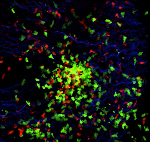
Video of Innate Immune Response Takes Top Prize in 2nd Annual Small World In Motion Competition

Posted on January 15, 2013
Nikon Instruments Inc. is pleased to announce the winners of the second annual Small World in Motion Photomicrography Competition. Top honors go to Dr. Olena Kamenyeva of the National Institute of Health’s NIAID - Laboratory of Immunoregulation. Dr. Kamenyeva’s video, titled “Sensing danger,” shows the immune response in the lymph node of a mouse. Kamenyeva says this video is representative of efficient innate immune reaction in the lymph node, which typically has been studied for the development of adaptive immune response.
In response to the exciting new trend in digital photomicrography of recording time-lapsed movies through the microscope, Nikon Small World in Motion was created as a sister competition under the Nikon Small World brand. Movies are judged on the merit of being visually outstanding as well as depicting the intersection of science and art.
The winning video perfectly demonstrates this delicate balance, as to capture this video, Dr. Kamenyeva needed to use some of the most advanced techniques available. The inguinal lymph node was imaged using a two-photon microscope equipped with an L25.0 x 0.95 water immersion objective. Together, this allowed for the visualization of actual events occurring in challenged lymphatic tissue.
Dr. Stefan Lüpold takes second place with a time-lapse movie showing sperm from two different males competing within the female reproductive tract of the fruit fly, Drosophila melanogaster. While competition between sperm is a widespread phenomenon throughout the animal kingdom - and a powerful evolutionary force driving species diversity - it has been nearly impossible to study the fundamental biological processes associated with such sperm competition occurring when sperm from different males mix inside of females. The very recent development of genetically-modified fruit flies that produce sperm with either green- or red-fluorescent heads (as seen in Lüpold’s movie) is now allowing scientists to answer important biological questions.
Dr. Nils Lindstrom earned third place honors with his video, “Growing complexity in the kidney.” Dr. Lindstrom’s research is focused on understanding how the kidney and nephrons are patterned during embryonic development. His image, captured as part of his ongoing research on that topic, shows a metanephric kidney, cultured in vitro and imaged over four days. Dr. Lindstrom submitted the time-lapse video because it’s such a striking example of how a kidney starts from a simple structure and gradually becomes a highly complex collecting duct system in a matter of days. He says that how tissues are structured and patterned is a fundamental aspect of kidney development.
“For the second year in a row, Nikon has received an incredible number of entries for Small World in Motion that are both visually mesmerizing and scientifically relevant, said Eric Flem, Communications Manager, Nikon Instruments. “Dr. Kamenyeva’s image is the perfect combination of cutting-edge science with aesthetics that we look for in Small World, to help raise the profile of science with scientists and non-scientists alike.”
Nikon Small World in Motion awarded three winners with a First, Second, and Third place prize, and recognized an additional 10 entries with Honorable Mentions. The full list of winners is as follows:
• 1st Place – Dr. Olena Kamenyeva, “Sensing danger”
• 2nd Place – Dr. Stefan Lüpold, “Sperm from two males competing within reproductive tract of a female fruit fly (Drosophila melanogaster)
• 3rd Place – Dr. Nils Lindstrom, “Growing complexity in the kidney”
• Honorable Mention – Dr. Oleg Lavrentovich, “Flowing liquid crystal”
• Honorable Mention – Mr. Andrew Dopheide, “Ciliates feeding on a bacterial biofilm”
• Honorable Mention – Mr. Michael Weber, “Action of the heart”
• Honorable Mention – Mr. Daniel von Wangenheim, “Arabidopsis endosomes”
• Honorable Mention – Mr. Wim van Egmond, “The rotifer Limnias melicerta”
• Honorable Mention – Mr. Fengzhu Xiong, “The making of the brain”
• Honorable Mention – Dr. Heiti Paves, “Onion bulb scale epidermis”
• Honorable Mention – Dr. Maria Nemethova, “CAR fish fibroblast”
• Honorable Mention – Ms. Phuong Anh Nguyen, “Microtubule asters recapitulated in a model cytoplasm”
• Honorable Mention – Ms. Kathryn Markey, “Broodstock Bay Scallop Argopecten irradians opening up to take a look around and feed”
The judges were Dominik Paquet of Rockefeller University and Michael W. Davidson, Director of the Optical and Magneto-Optical Imaging Center at the National High Magnetic Field Laboratory at Florida State University.
For additional information, follow the conversation on Facebook and Twitter @NikonSmallWorld.
