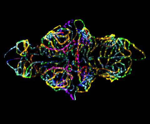
Blood-brain Barrier in a Live Zebrafish Embryo Takes First Place in 2012 Small World Competition

Posted on October 23, 2012
Nikon is pleased to announce the winners of the 2012 Small World Photomicrography Competition, with this year’s top honors going to Dr. Jennifer Peters and Dr. Michael Taylor of St. Jude Children’s Research Hospital. Their photomicrograph, “The blood-brain barrier in a live zebrafish embryo” is believed to be the first-ever image showing the formation of the blood-brain barrier in a live animal.
Nikon Small World recognizes excellence in photomicrography, honoring Drs. Peters and Taylor along with 97 other winners from around the world – some of whom won multiple times – who submitted images that showcase the delicate balance between outstanding scientific technique and exquisite artistic quality.
“Year over year, we receive incredible images from all over the world for the Nikon Small World Competition, and it is our privilege to honor and showcase these talented researchers and photomicrographers,” said Eric Flem, Communications Manager, Nikon Instruments. “We are proud that this competition is able to demonstrate the true power of scientific imaging and its relevance to both the scientific communities as well as the general public.”
First place winners Peters and Taylor partnered to capture the image highlighting their research of the blood brain barrier. “We used fluorescent proteins to look at brain endothelial cells and watched the blood-brain barrier develop in real-time,” said Drs. Peters and Taylor. “We took a 3-dimensional snapshot under a confocal microscope. Then, we stacked the images and compressed them into one – pseudo coloring them in rainbow to illustrate depth.”
The top five images this year come from a wide variety of artistic visual concepts and scientific disciplines who all share a common goal of outstanding photomicrographs that demonstrate superior technical competency and artistic skill.
Top Five Images:
1. Dr. Jennifer L. Peters and Dr. Michael R. Taylor, St. Jude Children’s Research Hospital; “The blood-brain barrier in a live zebrafish embryo”
2. Walter Piorkowski, “Live newborn lynx spiderlings”
3. Dr. Dylan Burnette, National Institutes of Health; “Human bone cancer (osteosarcoma) showing actin filaments (purple), mitochondria (yellow), and DNA (blue)”
4. Dr. W. Ryan Williamson, Howard Hughes Medical Institute (HHMI); “Drosophila melanogaster visual system halfway through pupal development, showing retina (gold), photoreceptor axons (blue), and brain (green)”
5. Honorio Cócera-La Parra, University of Valencia; “Cacoxenite (mineral) from La Paloma Mine, Spain”
This year’s judges were once again comprised of top science and media industry experts:
Daniel Evanko, Editor, Nature Methods; Martha Harbison, Senior Editor, Popular Science; Dr. Robert D. Goldman, Stephen Walter Ranson Professor and Chair, Department of Cell and Molecular Biology, Feinberg School of Medicine at Northwestern University and Liza A. Pon, Ph.D., Professor of Pathology and Cell Biology and Director, Confocal and Specialized Microscopy Shared Resource, Columbia University.
Top images from the 2012 Nikon Small World Competition will be exhibited in a full-color calendar and through a national museum tour. Follow the conversation on Facebook and Twitter @NikonSmallWorld.
