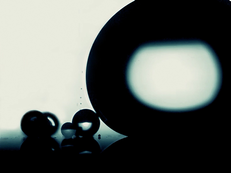
Coalescing Micro-Droplets Win The 10th Annual Nikon Small World in Motion Competition

First Place, 2020 Small World in Motion Competition: Internal flow dynamics of coalescing micro-droplets (~200x slower speed)
Posted on September 15, 2020
Nikon Instruments Inc. today unveiled the winners of the tenth annual Nikon Small World in Motion Photomicrography Competition. This year’s first place prize was awarded to Mr. Kazi Rabbi and Dr. Xiao Yan and for their eye-catching video of micro-droplets (made of 80% water and 20% ethanol) coalescing. Rabbi and Yan’s research at the University of Illinois at Urbana-Champaign is focused on creating surfaces that enhance the condensation and evaporation of liquid (repelling liquids from the surface they are on). People see condensation every day but are likely unaware of how enhancing condensation carries significant implications for sustainable living and creating energy efficient technology.
“Think about anything from keeping the pipes from freezing in winter to making your air conditioning unit run more efficiently,” said Rabbi, “If we can develop surfaces and materials that better repel liquids, we can create appliances, power systems, and other technologies that require less energy to run. It could lead to a more sustainable future.”
Rabbi and Yan conduct many experiments to see how liquids react to the functionalized surfaces (materials that can be used in the creation of new technologies) they have created, which is how they were able to capture this year’s top video. “Much of our microscopy is focused on visualizing how liquid droplets or condensate droplets interact with such surfaces at micro scale.” Yan said. This visualization is no easy feat to capture. The surface the droplets in the video are reacting to is one of Rabbi and Yan’s own design.
To capture the video, the pair used transmitted light microscopy. The biggest challenge, they said, was controlling the micro-droplet generation and growth. They had to use a frequency-controlled micro-droplet dispenser and a high-speed camera interfaced with a microscopic lens to accomplish the task – all while focusing on the perfect plane and maintaining perfect lighting. It was a two-person job.
“This year’s movies, and our winning video in particular, captures the spirt of Nikon Small World in Motion on the competition’s 10th anniversary,” said Eric Flem, Communications Manager, Nikon Instruments. “The winning video illustrates how highly sophisticated imaging techniques and systems can help us see and better understand common concepts as well as lead to improvements to technologies and products we all use in our everyday lives.”
Second place was awarded to Dr. Richard Kirby, a marine scientist focused on the study of plankton and their environments, for his darkfield video of a phoronid larva of a marine horseshoe worm. Dr. Kirby’s subjects can be very hard to capture because of their delicate nature. Despite their importance to many ecosystems, people know relatively little about these creatures, Kirby said. Microscopy, he continued, creates great images that attract the public's curiosity and assist in better understanding of their lives and the role they play in our ecosystems.
The 2020 judging panel included:
• Dr. Dylan Burnette, Assistant Professor of Cell and Developmental Biology at Vanderbilt University
• Dr. Christophe Leterrier, Group Leader at the Institute of Neurophysiopathology at CNRS and Aix-Marseille University
• Samantha Clark, Photo Editor at National Geographic
• Sean Greene, Graphics and Data Journalist at The Los Angeles Times
• Ariel Waldman, Chair of the External Council for NASA's Innovative Advanced Concepts Program
NIKON SMALL WORLD IN MOTION WINNERS
1st Place
Kazi Fazle Rabbi & Dr. Xiao Yan
University of Illinois at Urbana-Champaign
Mechanical Science and Engineering
Urbana, Illinois, USA
Internal flow dynamics of coalescing micro-droplets (~200x slower speed)
Transmitted Light
20x (Objective Lens Magnification)
2nd Place
Dr. Richard Ralph Kirby
The Plankton Pundit
Plymouth, Devon, United Kingdom
Planktonic larva of a marine horseshoe worm (Actinotrocha)
Darkfield
1.0x and 2.3x (Objective Lens Magnification)
3rd Place
Martin Kaae Kristiansen
My Microscopic World
Aalborg, Nordjylland, Denmark
A blackworm (Lumbriculus variegatus) displaying peristaltic movements
Polarized Light
4x (Objective Lens Magnification)
4th Place
Dr. Andrew Moore & Dr. Pedro Guedes-Dias
Howard Hughes Medical Institute (HHMI)
Janelia Research Campus
Ashburn, Virginia, USA
Fluorescent actin (Lifeact-EGFP) expressed in an embryonic rat hippocampal neuron
Confocal
100x (Objective Lens Magnification)
5th Place
Wojtek Plonka
Krakow, Malopolskie, Poland
Crystallization of a callus removal solution
Polarized Light
6.3X (Objective Lens Magnification)
HONORABLE MENTIONS
Dr. Gregory Adams Jr.
National Institute of Health
NHLBI
Bethesda, Maryland, USA
Morphing melanoma cells (alpha-Actintin shown in yellow; actin in red)
Confocal
60X (Objective Lens Magnification)
Massimo Brizzi
www.massimobrizzi.it
Empoli, Firenze, Italy
Colonies of rotifers with eggs
Darkfield
10X (Objective Lens Magnification)
Massimo Brizzi
www.massimobrizzi.it
Empoli, Firenze, Italy
Colonies of green algae (Volvox)
Darkfield
10X - 20X (Objective Lens Magnification)
Daniel Castranova & Dr. Brant Weinstein
NIH, NICHD
Section on Vertebrate Organogenesis
Bethesda, Maryland, USA
The first 22 hours of zebrafish development (blood vessels shown in green)
Confocal
4X (Objective Lens Magnification)
Dr. Douglas Clark
Paedia LLC
San Francisco, California, USA
Herb (Tradescantia spathacea) leaf stoma (breathing pore) responding to CO2 and RH transients
Brightfield, Image Stacking
50X (Objective Lens Magnification)
Dr. Stephan Daetwyler, Dr. Gloria Slattum, Dr. Jody Rosenblatt & Dr. Jan Huisken
UT Southwestern
Department of Cell Biology
Dallas, Texas, USA
Cancer cell metastasis in a developing zebrafish embryo
Light Sheet
10X (Objective Lens Magnification)
Frank Fox
Trier University of Applied Sciences
Konz, Rheinland-Pfalz, Germany
A stentor (ciliate) juggling green algae
Darkfield
20X (Objective Lens Magnification)
Frank Fox
Trier University of Applied Sciences
Konz, Rheinland-Pfalz, Germany
Hydra
Darkfield
10X (Objective Lens Magnification)
Karl Gaff
Dublin, Ireland
Oil droplets on a soap film
Reflected Light
4X (Objective Lens Magnification)
Ralph Claus Grimm
Jimboomba, Queensland, Australia
Ciliate (Prorodon viridis) showing its beating cilia and green zoochlorellae
Differential Interference Contrast (DIC)
60X (Objective Lens Magnification)
Roland Gross
Gruenen, Switzerland
Ciliates (Vorticella
sp. and Paramecium sp.)
Differential Interference Contrast (DIC)
10X (Objective Lens Magnification)
Eric Lind
Delmar, New York, USA
Developing freshwater snail embryo, inside the egg
Darkfield
4X, 10X and 40X (Objective Lens Magnification)
Rafael Martín-Ledo
IES Leonardo Torres Quevedo
Biología y Geología
Santander, Cantabria, Spain
Two larvae of a parasitic flatworm (Platyhelminthes)
Phase Contrast
10X (Objective Lens Magnification)
Rafael Martín-Ledo
IES Leonardo Torres Quevedo
Biología y Geología
Santander, Cantabria, Spain
A marine tardigrade (Batillipes lusitanus)
Phase Contrast
20X (Objective Lens Magnification)
Rogelio Moreno
Panama, Panama
Nematode
Polarized Light
10X (Objective Lens Magnification)
Rogelio Moreno
Panama, Panama
Ciliate (Frontonia)
Differential Interference Contrast (DIC)
20X (Objective Lens Magnification)
Andrei Savitsky
Cherkassy, Ukraine
Spathidium ciliate feeding on Vorticella ciliate
Phase Contrast
20X (Objective Lens Magnification)
Anjalie Schlaeppi
Morgridge Institute for Research
Department of Medical Engineering
Madison, Wisconsin, USA
Endocardium, cells lining the heart chambers, in a beating heart of a living, 2 day old zebrafish (Danio rerio)
Selective Plane Illumination Microscopy (SPIM)
16x (Objective Lens Magnification)
Dr. Wim van Egmond
Micropolitan Museum
Berkel en Rodenrijs, Zuid Holland, Netherlands
Cytoplasmic streaming in slime mold
Darkfield
10x (Objective Lens Magnification)
