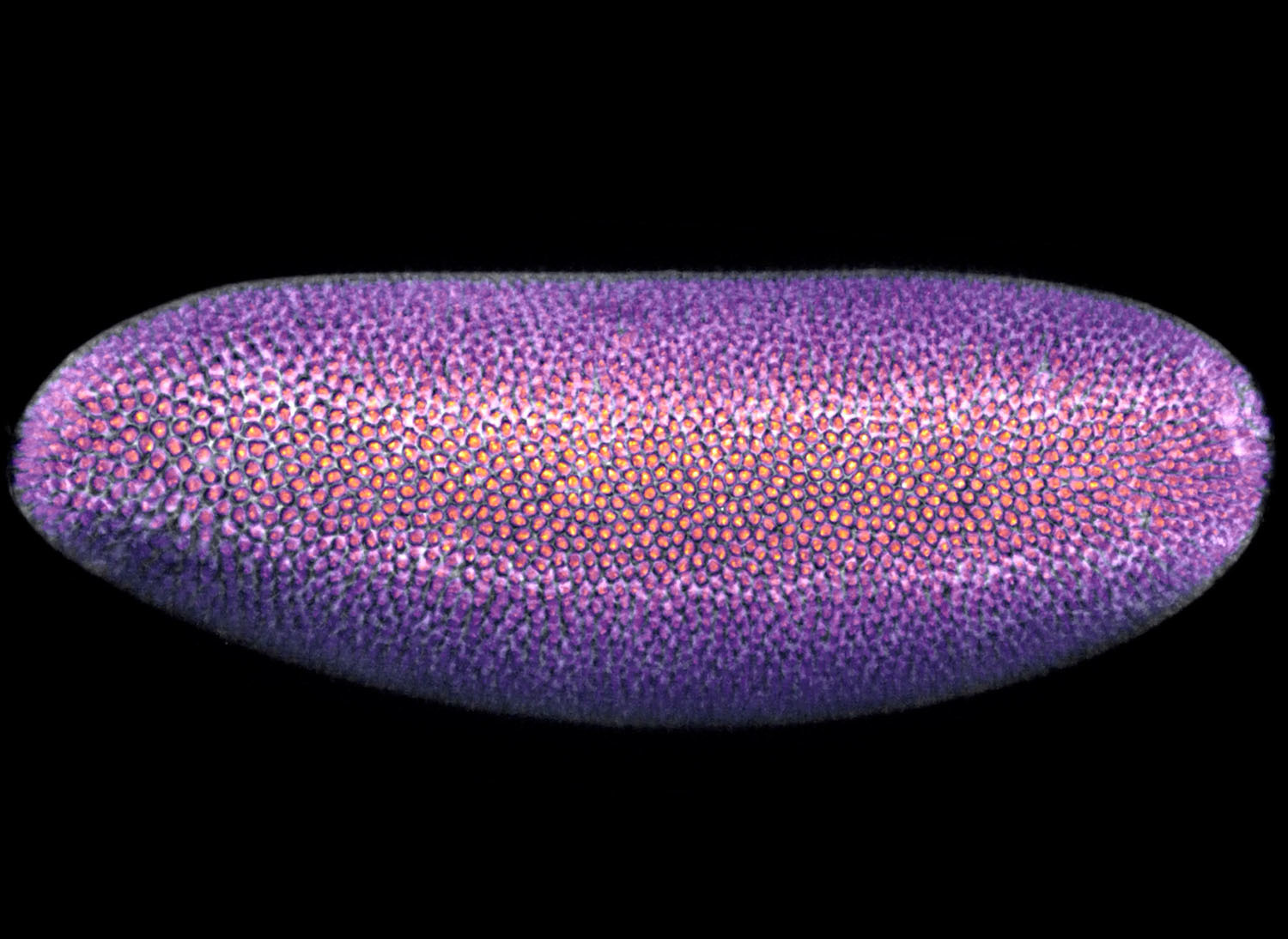
Nikon Instruments Inc. today unveiled the winners of its 14th annual Nikon Small World in Motion Video Competition, an integral component of the Nikon Small World competition, which is celebrating its 50th anniversary this year. The first-place prize was awarded to Dr. Bruno Vellutini of the Max Planck Institute of Molecular Cell Biology and Genetics for his video of mitotic waves in the embryo of a fruit fly (Drosophila melanogaster). The video reveals the dynamic processes of fly embryogenesis, crucial for uncovering genetic pathways that mirror those in humans and other mammals, with applications for cancer research, birth defects, and potential treatment development.
As a zoologist with a background in evolutionary and developmental biology, Dr. Vellutini is dedicated to advancing our understanding of how embryos build themselves from a single cell—a process fundamental to all animal life. His research, while focused on evolutionary questions, has broader implications for medical science, specifically rare neurological disorders and limb malformations in humans. “The beauty of basic research in biology,” says Dr. Vellutini, “is that what we learn in one organism is often applicable to others and has the potential to contribute to the understanding of human diseases.”
Dr. Vellutini’s winning video captures the rhythmic waves of division and tissue movements critical to proper embryonic formation in fruit flies. These processes are linked to mechanisms that can go awry, leading to the development of cancer and other diseases. For instance, during normal development, cells must organize precisely within tissues, a process maintained by cell-cell communication and mechanical feedback. Disruptions in these processes, such as the epithelial-mesenchymal transition—a process normal in embryogenesis but problematic when occurring unexpectedly—are known to contribute to the invasiveness of lung, liver, and breast cancer.
Dr. Vellutini adds depth to his video, saying, “Fruit fly embryos are in our homes, developing in our kitchens and our trash bins, are undergoing the same processes as shown in the video. I believe the video is particularly impactful because it shows us how these fascinating cellular and tissue dynamics are happening every day, all around us—even in the most mundane living beings.”
To capture this dynamic development, Dr. Vellutini used light sheet microscopy, a technique that allows for gentle imaging of live embryos while minimizing potential damage. “The biggest challenges I encountered were in mounting the embryos correctly and optimizing imaging conditions to capture clear, high-quality footage over an extended period. Balancing light exposure to avoid harming the samples was crucial,” Dr. Vellutini explained.
For the last 50 years, Nikon Small World has been a leading showcase of cutting-edge microscopy and artistic imaging. Since its inception in 2011, Small World in Motion has been a vital component of this gallery,” said Eric Flem, Senior Manager, Communications and CRM at Nikon Instruments. “As we enter a new era of the competition, we remain committed to highlighting the boundaries of innovation in scientific imaging. Nikon’s dedication to advancing science and art is especially evident in Dr. Vellutini’s winning entry, which stands as a testament to this legacy, capturing a mesmerizing movement within the microscopic world that helps deepen our understanding of a process that shapes life itself.”
Second place was awarded to Jay McClellan for his video of water droplets evaporating from the wing scales of a peacock butterfly (Aglais io). The final product used image stacking and a custom CNC motion control system to handle evaporating droplets and ensure smooth, rapid image capture.
Third place was awarded to Dr. Jiaxing Li for his video of an oligodendrocyte precursor cell in the spinal cord of a zebrafish.
The 2024 judging panel included:
- Adrian Coakley, Director of Photography at National Geographic Books
- Michelle S. Itano, Ph.D., Assistant Professor of Cell Biology and Physiology and Director of the Neuroscience Microscopy Core at the University of North Carolina at Chapel Hill
- Emily Petersen, Photography Managing Editor at Science Magazine
- Clare Waterman, Ph.D., Cell Biologist and Member of the National Academy of Sciences
- Jennifer C. Waters, Ph.D., Director of the Core for Imaging Technology & Education at Harvard Medical School
- Samantha Yammine, Ph.D., Neuroscientist and Science Communicator
NIKON SMALL WORLD IN MOTION WINNERS
1st Place
Dr. Bruno Vellutini
Max Planck Institute of Molecular Cell Biology and Genetics
Dresden, Saxony, Germany
Mitotic waves in the embryo of a fruit fly (Drosophila melanogaster)
Light Sheet
20X (Objective Lens Magnification)
2nd Place
Jay McClellan
Saranac, Michigan, USA
Water droplets evaporating from the wing scales of a peacock butterfly (Aglais io)
Image Stacking, Timelapse
5X (Objective Lens Magnification)
3rd Place
Dr. Jiaxing Li
Portland, Oregon, USA
An oligodendrocyte precursor cell in the spinal cord of a zebrafish
Confocal
20X (Objective Lens Magnification)
4th Place
Dr. Ignasi Vélez Ceron, Dr. Francesc Sagués & Dr. Jordi Ignés-Mullol
University of Barcelona
Department of Materials Science and Physical Chemistry
Barcelona, Spain
Friction transition in a microtubule-based active liquid crystal
Fluorescence
20X (Objective Lens Magnification)
5th Place
Quinten Geldhof
Winthrop, Massachusetts, USA
A baby tardigrade riding a nematode
Darkfield
10X (Objective Lens Magnification)
HONORABLE MENTIONS
Richard J. Albrecht
Altenstadt, Bavaria, Germany
Molting mayfly
Brightfield
2X (Objective Lens Magnification)
Thomas Barlow & Connor Gibbons
Columbia University
Department of Neurobiology and Behavior
New York, New York, USA
Spontaneous activity of pigment-filled chromatophores in the skin of the dwarf cuttlefish (Sepia bandensis)
Reflected Light
20X (Objective Lens Magnification)
Thomas Barlow & Connor Gibbons
Columbia University
Department of Neurobiology and Behavior
New York, New York, USA
Movement and chromatophore activity in a developing octopus embryo (Octopus hummelincki)
Darkfield, Stereomicroscopy
8X (Objective Lens Magnification)
Dr. Ailen Cervino
Baylor College of Medicine
Center for Precision Environmental Health (CPEH)
Houston, Texas, USA
Frog (Xenopus laevis) dorsal mesoderm cells
Confocal
40X (Objective Lens Magnification)
Dr. Luis Carlos Cesteros
Durango, Bizkaia, Spain
Algae (Synura uvella)
Differential Interference Contrast (DIC)
10X, 20X and 40X (Objective Lens Magnifications)
Nikky Corthout & Dr. Francesca Rizzollo
VIB
Center for Brain and Disease Research
Leuven, Vlaams-Brabant, Belgium
Melanoma cells showing mitochondria and lysosome dynamics
Confocal, Fluorescence
60X (Objective Lens Magnification)
Samantha Fallacaro, Dr. Apratim Mukherjee & Puttachai Ratchasanmuang
University of Pennsylvania
Department of Cell and Developmental Biology
Philadelphia, Pennsylvania, USA
Three views of a fruit fly (Drosophila melanogaster): adult, embryo, and histones tagged with GFP marking nuclei
Brightfield, Fluorescence, and Light Sheet
10X-100X (Objective Lens Magnifications)
Dr. KrassiMira A. Garbett & Richard Sando
Vanderbilt University
Department of Pharmacology
Nashville, Tennessee, USA
Live mouse neurons with fluorescently labeled points of connection (7 hour time-lapse)
Confocal
60X (Objective Lens Magnification)
Quinten Geldhof
Winthrop, Massachusetts, USA
Mosquito larva feeding
Darkfield
4X (Objective Lens Magnification)
Dr. Saikat Ghosh
National Institutes of Health
NICHD
Bethesda, Maryland, USA
Dynamic trafficking of mitochondria and lysosomes along microtubule tracks of neuronal highways
Confocal
63X (Objective Lens Magnification)
Cora A. Harris
Charlotte, North Carolina, USA
Crystallization of magnesium sulfate (MgSO4) salt crystals
Polarized Light
10X (Objective Lens Magnification)
Bre Hewitt
Drexel University
Department of Biology
Philadelphia, Pennsylvania, USA
Human fibroblast climbing a 3D 'rope' of extracellular matrix (gray) by squeezing the nucleus (cyan) through tight spaces. Actin cytoskeleton (orange).
Confocal, Fluorescence
60X (Objective Lens Magnification)
Dr. Patrick Colin Hickey
NIPHT LIMITED
Edinburgh, Midlothian, United Kingdom
Dynamics of hyphal growth, cytoplasmic flow and movement of nuclei within a colony of filamentous fungus (Neurospora crassa). Time elapsed: 1.5 hours
Confocal
40X (Objective Lens Magnification)
Dr. Alvaro Migotto
Centro de Biologia Marinha
São Sebastião, São Paulo, Brazil
Parchment worm larva eating a copepod
Darkfield
4X (Objective Lens Magnification)
Dr. Andrew Moore
Howard Hughes Medical Institute
Janelia Research Campus
Ashburn, Virginia, USA
Crawling tissue culture cell
Confocal
63X (Objective Lens Magnification)
Dr. Andrew Moore
Howard Hughes Medical Institute
Janelia Research Campus
Ashburn, Virginia, USA
Time-lapse of cultured cells crawling and exploring
Confocal
63X (Objective Lens Magnification)
Rogelio Moreno
Panama, Panama
Sea angel (Pterapod sp.)
Darkfield
4X (Objective Lens Magnification)
Irina Petrova Adamatzky
University of the West of England Bristol
College of Arts, Technology and Environment
Bristol, Somerset, United Kingdom
Malaysian flower mantis (Creobroter urbanus)
Reflected Light
5X (Objective Lens Magnification)
Benedikt Pleyer
Kirchberg, Bavaria, Germany
White blood cells in menstrual blood consuming uterus mucosa cells
Differential Interference Contrast (DIC)
60X (Objective Lens Magnification)
Benedikt Pleyer
Kirchberg, Bavaria, Germany
Chemical reactions within a single drop of water
Darkfield
25X - 50X (Objective Lens Magnifications)
Catherine Porter, Aria Huang, Hunter Nichols & Dr. Alex Hughes
University of Pennsylvania
Department of Bioengineering
Philadelphia, Pennsylvania, USA
Time-lapse of canine kidney cells, precisely arranged by photopatterned DNA tethers, creating a replica of Vermeer's painting, “Girl with a Pearl Earring”
Confocal, Fluorescence
4x (Objective Lens Magnification)
Jan Rosenboom
Rostock, Mecklenburg Vorpommern, Germany
Ignition of an arc lighter
Darkfield, Reflected Light
4X (Objective Lens Magnification)
Jennifer Silverman
Vanderbilt University
Cell & Developmental Biology
Nashville, Tennessee, USA
Two HeLa cells expressing an actin binding protein generate large numbers of dynamic protrusions, which are evident in the space between the cells
Confocal
100X (Objective Lens Magnification)
Sebastian Sparenga
McCrone Research Institute
Chicago, Illinois, USA
Dinitronaphthalene (a chemical compound) recrystallizing upon cooling
Polarized Light
4X (Objective Lens Magnification)
Sanjay Sunil Kumar & Steven Reger
Schulte-Merker Lab, Institute for Cardiovascular Organogenesis and Regeneration
University Hospital, Muenster, Germany
Muenster, NRW, Germany
Blood endothelial cells forming a lumen during zebrafish development
Confocal
63X (Objective Lens Magnification)
Dr. Wim van Egmond
Micropolitan Museum
Berkel en Rodenrijs, Zuid Holland, Netherlands
Starfish gastrula, escaping from the egg
Darkfield, Differential Interference Contrast (DIC)
16X (Objective Lens Magnification)
Dr. Daniel Wehner & Nora John
Max Planck Institute for the Science of Light
Department of Biological Optomechanics
Erlangen, Bavaria, Germany
Immune response (neutrophils, cyan; macrophages, orange) to spinal cord injury in zebrafish
Confocal
10X (Objective Lens Magnification)
Dr. Cara Winter
Duke University
Department of Biology
Durham, North Carolina, USA
17-hour time-lapse of a growing thale cress (Arabidopsis thaliana) root showing induction of a gene that controls stem cell division, SHR (green), nuclei (red)
Confocal
40X (Objective Lens Magnification)
Chew Yen Fook
Woodend, Waimakiriri, New Zealand
Oligochaete worm (Chaetogaster sp.) feeding on a water flea (Chydrorus sp.)
Darkfield, Polarized Light
4X (Objective Lens Magnification)
Wenting Zhu
Beauty of Science
Hefei, Anhui Province, China
Electrodeposition of silver on copper
Reflected Light, Stereomicroscopy
2.5X (Objective Lens Magnification)
Wenting Zhu
Beauty of Science
Hefei, Anhui Province, China
Convection cells in a paint-alcohol mixture
Reflected Light, Stereomicroscopy
5X (Objective Lens Magnification)
To view all the winners, click here.