In this article, you’ll meet the members of the 2024 judging panel who reviewed thousands of microscopy submissions from across the globe to select this year's winners. What makes this year even more extraordinary? It marks the 50th anniversary of this incredible competition, celebrating half a century of exhibiting the mesmerizing beauty of microscopic imagery. Join us in commemorating this golden milestone as we honor this year’s judges!
Meet the JudgesMeet the Judges
Meet the 2024 Judging Panel
Nikon Small World showcases brilliant imagery and artistic feats in science, and the unseen world around us. With every new year, the Nikon Small World judging panel plays an integral role in selecting the images and videos that best blend science and artistry. Nikon strives to select a panel that exemplifies a range of expertise – a team that can evaluate a photo or video based on technical prowess, scientific significance, aesthetic appeal, and power to convey a story to the viewer.
Adrian Coakley
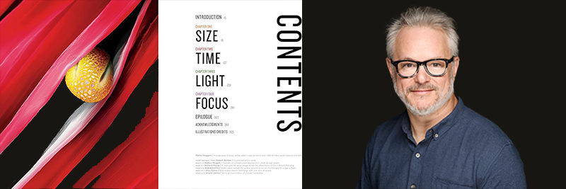
From the pages of “Invisible Wonders,” a book Adrian Coakley helped bring to life
For the past 136 years, National Geographic has been renowned for its vivid articles on the world's geography, history, nature, science, and culture. Adrian Coakley has been instrumental in maintaining that legacy, playing a major role in bringing this groundbreaking vision to life.
Since 2007, Coakley has been immersed in a world of stunning imagery, working on projects that explored everything from the mysteries of outer space to the origins of humanity. His talent for capturing the essence of adventure soon led him to National Geographic Magazine, where he brought these themes to both print and digital formats.
Coakley’s journey has been nothing short of compelling. In 2019, he took on the role of Director of Photography for National Geographic Books. His passion for visual storytelling then took center stage last year when he teamed up with renowned science photographer Anand Varma to develop the captivating coffee table book, “Invisible Wonders.”
Spanning 368 pages, the book reveals a world often unseen by the naked eye, from the microscopic to the cosmic. Reflecting on the experience, Coakley remarked, “Working with Anand and helping to structure and communicate his vision for the book was one of the highlights of my career. Anand’s fascination with how the camera can reveal hidden wonders, sometimes quite literally under our noses, perfectly encapsulates how science, art, and photography converge to illuminate the awe-inspiring beauty of our world.”
For Coakley, the intersection of science and art isn’t just a professional focus; it’s a passion that drives him, which made him a perfect candidate for the 2024 Nikon Small World judging panel. His extensive career with National Geographic gave him a profound appreciation for the intricate beauty captured in microscopic imagery. “Bridging art and science is crucial because it deepens our understanding of the world and if we’re lucky, ourselves. By highlighting the connections between the largest and smallest components of our world, we can open viewers' eyes to some of the intricacies of life and our natural world.”
"I enjoyed being on a panel with a great mix of scientists, science communicators, and the fantastic Nikon team,” said Coakley. “Despite lacking a science background, I was truly honored to participate in the judging panel for Nikon Small World, especially in its 50th anniversary year. The work was amazing, and seeing the global range of creative projects was truly inspiring.”
In the fall, Coakley’s team will release a book titled, "Infinite Cosmos”, on the James Webb Telescope, as well as another photography book titled, “World From Above,” curated by photographer Jeff Kerby.
Dr. Michelle S. Itano
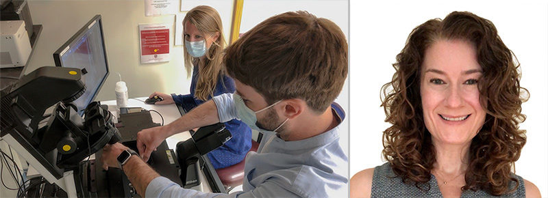
Dr. Michelle S. Itano welcoming 60 STAR (Scientific Training and Research) program students to experience life in a research lab and meet real scientists
From unearthing hidden realms to pioneering breakthrough scientific methods, Dr. Michelle S. Itano isn’t just advancing the scientific field—she’s redefining it. With over 16 years of experience, she developed and applied innovative fluorescence microscopy techniques to answer critical and previously intractable questions in cell and neurobiology. Her career began with a focus on genetic and genomic research using fruit flies, which laid the foundation for her fascination with microscopy.
During her graduate studies with Dr. Ken Jacobson at the University of North Carolina (UNC) School of Medicine, she embraced super-resolution microscopy to dive deep into how proteins in cell membranes play a role in human diseases.
Her postdoctoral work with Dr. Sanford Simon at The Rockefeller University allowed her to merge genetics, cell biology, virology, and biophysics, further enhancing her skills in advanced imaging techniques. “Integrating different scientific disciplines and pushing the boundaries of microscopy was an exhilarating challenge,” Dr. Itano shared. “It was an incredible opportunity to advance our understanding of complex biological processes.”
Now, as the director of the Neuroscience Microscopy Core and assistant professor of Cell Biology & Physiology at UNC-Chapel Hill, Dr. Itano is dedicated to training and consulting with researchers on advanced imaging.
She’s also been instrumental in improving data accessibility and fostering collaboration, such as with the development of an Omero server for microscopy data. She’s passionate about making science engaging, noting, “I believe it’s crucial that research technologies and results are shared with the public. Art and science together can make complex discoveries both captivating and understandable.”
Dr. Itano’s career pays homage to a lifelong passion for exploring how things work, stemming from her experience as a third-generation research scientist and her early days in Boulder, Colorado. Her role as a judge in science and art competitions highlights her commitment to blending science with visual expression. “Art allows us to communicate beyond language,” she noted. “It helps to bridge the gap between intricate scientific concepts and the public, making the wonders of science both accessible and inspiring.”
Reflecting upon her recent experience on the 50th anniversary panel, Dr. Itano added, “I loved the entire experience! From viewing the impressive array of submissions, to learning about the long history and impact of the Small World contest, to hearing more from the other judge's perspectives and expertise, it was truly a celebration of science and curiosity.”
She went on to say, “Getting to learn more about Mike Davidson and his legacy was very important to me – and timely – as an organization I work with, BioImaging North America, has just established the Michael W. Davidson Memorial Award which will be given for the first time this coming fall.”
Emily Petersen
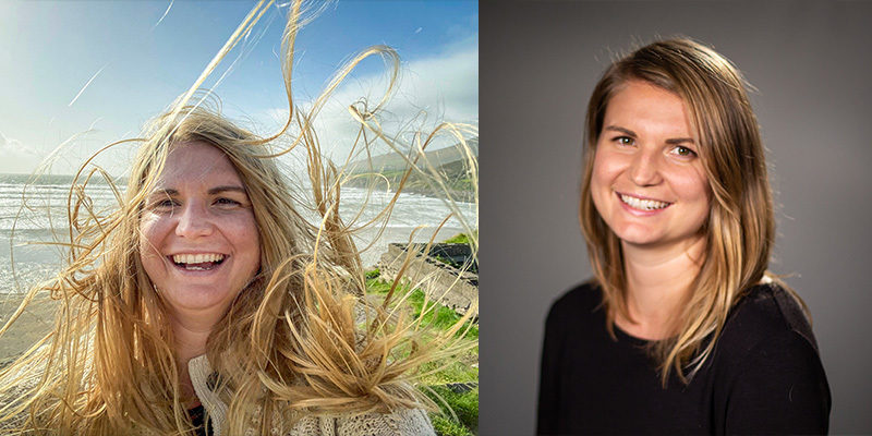
Emily Petersen enjoying the Inch Beach in Ireland
Emily Petersen’s career path to becoming the Photography Managing Editor at Science magazine is anything but conventional – as she puts it, “I used the side door.” After studying biology at Virginia Tech and working in a histology lab, she shifted gears to explore her passion for visual storytelling.
She combined environmental science, film, and photography in her master’s degree at American University, which led to an internship at Science in 2015. Emily has since moved from photo editing to managing the photo team, where she turns complex scientific concepts into captivating visuals.
Petersen’s blend of science and art stems from her fascination with documentaries like Planet Earth. It dawned on her that while scientists have deep knowledge, they often need help communicating it effectively. “I realized that scientists know so much about the world yet often need help explaining it into something compelling for those who never studied it,” she shared. For Petersen, great visuals are about more than technical skill—they need originality and a strong message. “Whenever I take a picture or video, the phrase that runs through my head is: ‘Is it interesting?’”
Judging competitions like Nikon Small World has been a blast for Petersen, who enjoys the diverse perspectives of her fellow judges. “It generated fascinating discussions when it came to advocating and defending the imagery,” she noted.
She sees the fusion of science and art as a way to engage and educate the public, combining logical learning with emotional impact. “Great art can make you care about something you knew nothing about,” she adds. While she’s keeping her upcoming projects under wraps, Emily’s expertise in selecting compelling images continues to shine.
You can find Petersen’s work on Science and their Visuals Blog.
Dr. Clare Waterman
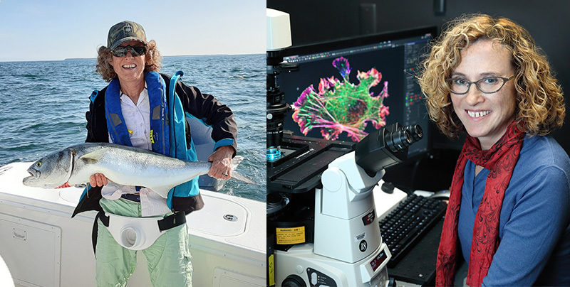
Dr. Clare Waterman enjoying a fishing adventure
Our most avid fans may recognize Dr. Clare Waterman – a celebrated cell biologist and veteran judge for the Nikon Small World Competition, she is known for her sharp wit and keen eye in identifying stunning images. With nine competitions under her belt, she masterfully evaluates intricate scientific techniques and cutting-edge imaging in her role as a judge.
Reflecting on her journey, Dr. Waterman shared, "Judging the Nikon Small World Competition over the years has been a true highlight, especially being part of the 50th anniversary. It's incredible to witness the blend of art and science through these remarkable images."
Dr. Waterman's 25-year career in imaging and microscopy began with a B.A. in biochemistry from Mount Holyoke College and an M.S. in exercise science from the University of Massachusetts. Initially an aspiring strength trainer, she was captivated by electron microscope images of muscle, which eventually steered her toward cell biology and microscopy.
After earning her Ph.D. in cell biology from the University of Pennsylvania in 1995, Dr. Waterman spent nine years as a professor at the Scripps Research Institute. Her career has been dedicated to imaging living movement at the microscopic scale, unveiling the dynamic interactions within cells.
Dr. Waterman truly enjoys judging the Nikon Small World Competition, where she gets to admire the thoughtful work created by her fellow scientists. Her cutting-edge work, including groundbreaking discoveries about how organelle systems interact within cells, highlights her deep expertise and passion for both microscopy and visual storytelling.
When she's not in the lab, you can find Dr. Waterman fishing in Woods Hole, Massachusetts.
Dr. Jennifer C. Waters
Students participating in the novel fellowship program created by Dr. Jennifer C. Waters.
Dr. Jennifer C. Waters has navigated an impressive career path, blending her expertise in microscopy with a passion for education. After earning her Ph.D. in biology from UNC-Chapel Hill where she focused on quantitative fluorescence live-cell imaging, Dr. Waters joined Harvard Medical School in 2001. As Director of the Core for Imaging Technology & Education, she trains scientists in light microscopy techniques and creates online resources such as the Microcourses YouTube channel and Microtutor – a pursuit she finds both fulfilling and exhilarating.
Dr. Waters’ scientific curiosity was sparked early by her father’s telescope hobby. “We would set up his telescopes on a local golf course and capture images of the sky, which fascinated me,” she recalled. This early fascination, combined with inspiring high school teachers, guided her toward a career that combines scientific precision with educational outreach.
For Dr. Waters, a standout microscopy image or video seamlessly merges technical skill with visual allure. “A compelling image or video in microscopy combines technical expertise with an element of beauty or interest that captures the viewer’s attention,” she noted. Her role as a judge has been enlightening, and she was especially pleasantly surprised by the high volume of entries.
She values the intersection of science and art, finding it crucial for making complex concepts accessible and engaging. With her background in microscopy and education, Dr. Waters’ insights as a judge were critical in identifying the winners of the monumental 50th anniversary competition.
Outside of work, she enjoys sewing and nurturing her collection of over 100 houseplants, which provide her with creative relaxation.
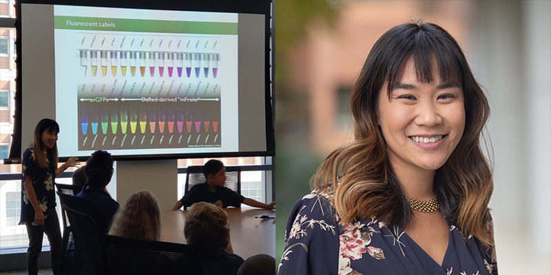
Dr. Samantha Yammine

Dr. Samantha Yammine’s career is a brilliant fusion of brain science and visual storytelling. With a Ph.D. from the University of Toronto where she explored neural stem cells, Samantha now dazzles audiences as a science communicator.
Through social media, TV, and radio, she turns complex scientific concepts into engaging and accessible content for the general public. She’s a regular science expert on CTV’s The Good Stuff with Mary Berg and has made guest appearances on various programs on Netflix, TVO Kids, CBC GEM, Discovery UK, CBC Radio, Seeker, and AsapSCIENCE.
Her love for blending science with art began in the lab. “When I finally got to step foot in a lab, it was a mesmerizing sight,” she recalled. Sharing captivating lab photos online revealed the beauty of science, sparking her passion for making science visually compelling.
Dr. Yammine finds that the real allure of an image or video lies in its context. “While of course blank space, focused lines, and strong colors can make an image or video pop, it's the story behind the image or video that captivates me. When we can make the connection between what we're seeing and the impact it can have on our world, that's when I really become obsessed,” she explained. She’s thrilled by the novelty of seeing familiar concepts in striking new ways, and as a judge, she was particularly impressed by the high-quality submissions and the challenge of capturing living cells in action.
Judging was a highlight, allowing her to celebrate the intersection of science and art with fellow experts. “Being surrounded by scientists and photojournalists with an expert eye for art and imagery was energizing,” she said. “Beyond the incredibly fun experience of judging, I also just love that we got to get together to celebrate science and art. There are very few opportunities to do that at such a high level, so it is a genuine privilege to now be a part of this legacy.”
For updates on her exciting projects, follow her on Instagram, TikTok, and Threads @science.sam.
The Nikon Small World in Motion video winners will be announced in September, and winners of the Small World photomicrography competition will be released in October.
To stay up to date with the "Meet the Judges" series and receive your daily dose of Nikon Small World, follow us on Instagram, Twitter, and Facebook. Be sure to follow Nikon Instruments for the latest updates on equipment and technology.