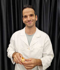
For Dr. Alexandre Dumoulin, the first time he used a microscope significantly transformed his perspective on organism development – the opportunity to witness intricate details of intact cells, particularly within their natural environment, deeply inspired him to embark on a career in academic research.
Masters of MicroscopyMasters of Microscopy

Dr. Alexandre Dumoulin on Exploring the Complexities of Neurodevelopmental Disorders
Welcome to Masters of Microscopy: The People Behind the Lens, where we showcase and celebrate the individuals who are the heart of the Nikon Small World competitions. They are scientists, artists, researchers, educators, and everyday curious individuals who uncover the fascinating microscopic world around us.

Dr. Alexandre Dumoulin
His passion to explore the depths of biological processes and contribute to the scientific understanding of the natural world, led him to earn a bachelor's and a master's degree in biochemistry at the University of Geneva. In 2013, Dumoulin moved to Germany to complete his Ph.D. through the Max Delbrück Center for Molecular Medicine in the Helmholtz Association.
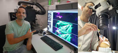
Finally, Dumoulin joined the lab of Professor Esther Stoeckli with the Department of Molecular Life Sciences at the University of Zurich as a postdoctoral researcher in December of 2017. Since then, he has explored how axons are guided towards their targets in developing central nervous systems.

Dumoulin conducting research in the Department of Molecular Life Sciences at the University of Zurich
“My research focuses on investigating the developmental processes of neurons in chick and mouse embryos,” said Dumoulin. “By studying these organisms, I aim to enhance our comprehension of how the nervous system functions and identify potential factors contributing to neurodevelopmental disorders.”
This year, Dumoulin wowed the Nikon Small World judges with his 48-hour time-lapse video of developing neurons connecting to the opposite side of the central nervous system in a chick embryo. While beautiful, Dumoulin’s video plays a significant role in understanding the potential deviations in neurodevelopmental disorders that occur in the central nervous system, such as autism spectrum disorder and schizophrenia.
Dumoulin’s 48-hour time-lapse video of developing neurons connecting to the opposite side of the central nervous system in a chick embryo
Neurons are responsible for carrying information throughout the human body. They are connected with long extensions known as axons and these axons traverse the nervous system before eventually forming synapses. Dumoulin’s video showcases these lengthy axons projecting across the midline, which serves as a boundary between the two hemispheres of the central nervous system. In neurological disorders, axons are impaired and unable to make their intended journeys.
“The nervous system is an immensely complex and intricate system composed of a myriad of units that are connected to one another,” said Dumoulin. “In this video, we see single units and how they behave.”
To capture the video, Dumoulin applied a new imaging method to visualize the live transfer of information from cells. “The biggest challenge was to discover a feasible method to access these neurons and capture images over an extended period of time,” said Dumoulin. “A combination of precise dissection skills and adapted microscopy techniques proved to be the key.”
In Dumoulin’s eyes, the competition provides an opportunity to share his research efforts and passion for microscopy with the world, “I wanted to share these mesmerizing developing neurons with the public. To me, that's the essence of this competition, highlighting the beauty of nature through the lens of scientific research.”
Outside his academic research, Dumoulin has practiced the Brazilian martial art, capoeira, since 2009 and continues to perfect his movements.

Dumoulin practicing capoeira
To stay up to date with the "Masters of Microscopy" series and receive your daily dose of Nikon Small World, follow us on Instagram, Twitter, and Facebook. Be sure to follow Nikon Instruments for the latest updates on equipment and technology.