Armed with education in marine biology and a passion for aquatic creatures, Daniel Castranova found his way to becoming a master of microscopy through his career at the National Institutes of Health. He started his career by caring for and managing the husbandry of the zebrafish in Dr. Brant Weinstein’s lab at the Eunice Kennedy Shriver National Institute of Child Health and Human Development, part of the NIH. Through his work, Castranova began assisting with the photography of the lab’s zebrafish and developed an affinity for capturing beautiful images from these tricky-to-photograph live specimens under the microscope. Over the years, he found a passion for imaging these tricky-to-capture creatures and has become an expert at creating stunning visuals to accompany the study of these fish.
Masters of MicroscopyMasters of Microscopy

What Zebrafish Teach Us About Cancer and Alzheimer’s Drug Therapies
Welcome to Masters of Microscopy: The People Behind the Lens, where we showcase and celebrate the individuals who are the heart of the Nikon Small World competitions. They are scientists, artists, researchers, educators and everyday curious individuals who uncover the fascinating microscopic world around us.

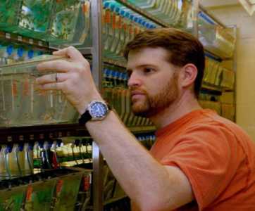
Castranova attends to his zebrafish at Dr. Brant Weinstein’s laboratory
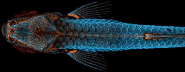
Castranova’s years of study and hard work culminated in this year’s top prize at NSW, which depicts his artfully rendered and technically immaculate photo of a juvenile zebrafish. The image is a dorsal view of the head of a fish with fluorescently “tagged” skeleton, scales (blue) and lymphatic system (orange), taken using confocal microscopy and image-stacking.

Castranova’s winning image; taken with the assistance of teammate Bakary Samasa
“The image is beautiful, but also shows how powerful the zebrafish can be as a model for the development of lymphatic vessels,” Castranova said, “Until now, we thought this type of lymphatic system associated with the nervous system only occurred in mammals. By studying them, the scientific community can expedite a range of research and clinical innovations – everything from drug trials to cancer treatments. This is because fish are so much easier to raise and image than mammals.”
To create this single stunning visual, Castranova stitched together more than 350 individual images. The image was acquired using a spinning disk confocal, merging together maximum intensity projections of three separate image Z stacks to generate the final reconstructed image. Crucially, it was taken as part of an imaging effort that helped Castranova’s team make a groundbreaking discovery - zebrafish have lymphatic vessels inside their skull that were previously thought to occur only in mammals. Their occurrence in fish, a much easier subject to raise, experiment with, and photograph, could expedite and revolutionize research related to treatments for diseases that occur in the human brain, including cancer and Alzheimer’s.
Castranova explains further the significance of the discovery. “Whether it's conducting drug screens more quickly, examining how immune cells behave or understanding how different molecules make vessels grow more, these are all important factors for cancer treatments.”
Discovery -- years in the making.
Daniel’s early academic work laid a foundation for his research interests and area of study. His undergraduate work focused on marine science and biology -- followed by a master’s degree in animal science from the University of Maryland, College Park. Daniel has spent the past sixteen years working for Charles River as a research technician in the Weinstein Laboratory at the Eunice Kennedy Shriver National Institute of Child Health and Human Development at the National Institutes of Health in Bethesda Maryland. The lab is focused on the study of vascular development, or the formation of the elaborate networks of vessels that carry blood (circulatory system) or lymph (lymphatic system) through the body
He also co-manages the microscopes for the Division of Developmental Biology with responsibilities including training new users, maintaining systems, and assisting other researchers with image acquisition. This experience would lend itself well to creating his winning image and many other stunning visuals of zebrafish.

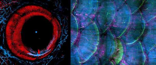
An eye of an adult zebrafish (left) and an image of zebrafish scales (right) both taken by Castranova.
For those interested in pursuing microscopy, Castranova recommends tapping into any resource available to you, like colleagues or certification courses. He found the Quantitative Fluorescent Microscopy course at Mount Desert Island particularly valuable. But like with most skills, practice and patience makes perfect. “I’ve spent hours and hours imaging zebrafish,” he says, “it takes practice to get it just right. That said, I am self-taught. I’ve taken a few courses, but a lot of what I learned came from observing others and putting the time in myself.”
Visualizations are so important, he says, because they can “really spark people’s curiosity and interest in important scientific issues.”
A passion since childhood.
When Daniel was a young boy, his father used to take him to visit the mountains and lakes of northern New Jersey to enjoy pan fishing. Ever since then, Castranova always had an interest in fish and aquatic animal life, which helps to explain why he pursued research and academic pursuits in marine biology and animal science. Today, he enjoys taking his own children fishing and out exploring nature.

Castranova fishes with his children
“I thought maybe I would be out in the field, on a boat, not necessarily in a lab. But I like the job that I have. I still get to explore nature and peak into unseen worlds and share those findings with others.” said Casanova.
We look forward to seeing what is next from this exceptional scientist who has already taught us so much about the small worlds that surround us.
To stay up to date with the "Masters of Microscopy" series and receive your daily dose of Nikon Small World, follow us on Instagram, Twitter, and Facebook. Be sure to follow Nikon Instruments for the latest updates on equipment and technology.