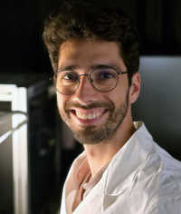
Hailing from São Paulo, Brazil, Dr. Bruno Vellutini’s fascination with biology was sparked at an early age, when a visit to a marine station and an encounter with an octopus left a lasting impression. Growing up snorkeling in the coastal waters and playing with toy microscopes, he has always been drawn to the wonders of the natural world. This curiosity soon evolved into a career that would take him across continents, from a bustling marine laboratory in Brazil to cutting-edge research institutes in Norway, and finally to the Max Planck Institute of Molecular Cell Biology and Genetics in Germany.
Masters of MicroscopyMasters of Microscopy
Dr. Bruno Vellutini on Decoding Life's Blueprint
Welcome to Masters of Microscopy: The People Behind the Lens, where we showcase and celebrate the individuals who are the heart of the Nikon Small World competitions. They are scientists, artists, researchers, educators, and everyday curious individuals who uncover the fascinating microscopic world around us.
At the heart of Dr. Vellutini’s work is a fundamental question: How does a single cell give rise to the complex forms and functions of a living organism? It’s a question that has driven Dr. Vellutini to explore the evolution of embryonic development, studying how the smallest of creatures—like fruit flies—can reveal the most profound biological insights, with far-reaching implications. While his primary focus is on evolutionary science, Dr. Vellutini’s research offers critical insights that serve to better our understanding of human diseases such as cancer and birth defects, where human developmental pathways are similar to those in the animals he studies.
His first-place video is a window into the rhythmic dance of cell division and tissue organization that transforms a group of nuclei into a fully formed embryo. The video begins at the tenth cycle of nuclear division and follows the synchronized waves of mitosis as they cascade through the embryo. As the cells organize and membranes form, the embryo begins to take shape.
Dr. Vellutini’s video of mitotic waves in the embryo of a fruit fly (Drosophila melanogaster)
“After the embryo is fertilized, the genetic material undergoes multiple rounds of division, creating many nuclei in a shared cytoplasm, known as the syncytium. These nuclei divide synchronously, causing rhythmic waves visible in the video,” Dr. Vellutini explains. “After 14 cycles of division, the nuclei become distributed around the embryo's surface. Following this, the embryo undergoes a dramatic phase called gastrulation, where the tissues move and reorganize, leading to significant morphological changes. This complex process involves multiple parts of the tissue folding simultaneously like origami, which is particularly noticeable at the embryo’s tail and neck regions.”
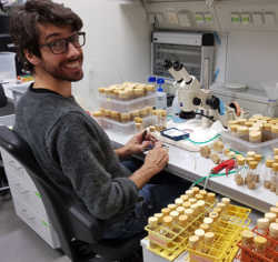
Dr. Vellutini tending to fly stocks at the Max Planck Institute of Molecular Cell Biology and Genetics
The significance of this work extends far beyond the realm of flies. “Many of the genetic pathways we study in flies are remarkably similar to those in humans,” Dr. Vellutini emphasizes. “Discoveries made in these tiny organisms can have direct applications in understanding diseases like cancer, which can occur when the pathways are disrupted.” For example, epithelial-mesenchymal transition, a process crucial in embryonic development, is also involved in the progression of different cancers such as lung, liver, and breast cancer. Additionally, signaling pathways like the Hedgehog pathway, first elucidated in fruit flies, are pivotal in understanding and treating conditions like basal cell carcinoma in humans.
Capturing such intricate details is no small feat. Dr. Vellutini’s mastery of light sheet microscopy—a technique that allows for gentle, live imaging of embryos—has been instrumental in his success. The process requires a delicate balance of preparation, from carefully mounting the embryos to optimizing the imaging conditions.
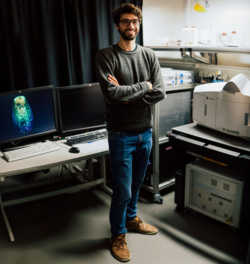
Dr. Vellutini with a light sheet microscope, taken by Magdalena Gonciarz for the Portrait of Science
Dr. Vellutini feels a deep sense of honor in being part of the storied tradition of Nikon Small World as the competition celebrates its 50th anniversary. “I truly value how this competition bridges the gap between scientists, artists, and the public, making complex concepts more accessible and engaging. It provides a special opportunity for everyone to admire the beauty of the microscopic world while also showcasing significant strides in scientific research.”
Looking ahead, Dr. Vellutini’s excitement for the future is palpable. He’s branching out, comparing developmental processes across different species of flies, and exploring how mechanical forces within tissues might influence embryonic evolution. His work continues to push the boundaries of what we know about life’s most fundamental processes, with each new project bringing us closer to understanding the delicate balance of forces that shape the natural world. Furthermore, he hopes that some of his future findings will contribute to understanding human disorders, such as those linked to genes like FOXG and EVX, which are associated with rare neurological conditions and limb malformations.
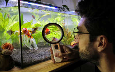
Beyond his research, Dr. Vellutini has several hobbies that balance his intense scientific work with creative and relaxing pursuits. When he’s not peering through the microscope, he can be found tending to his planted aquariums or playing the bass guitar. He is currently recording his own biology-inspired, bass-oriented electronic music project called Germband.
You can find Dr. Vellutini’s website here.
To stay up to date with the "Masters of Microscopy" series and receive your daily dose of Nikon Small World, follow us on Instagram, Twitter, and Facebook. Be sure to follow Nikon Instruments for the latest updates on equipment and technology.