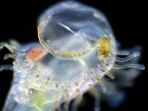
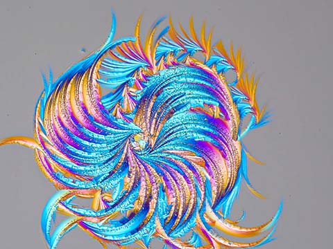
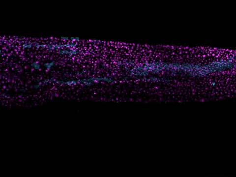
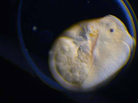
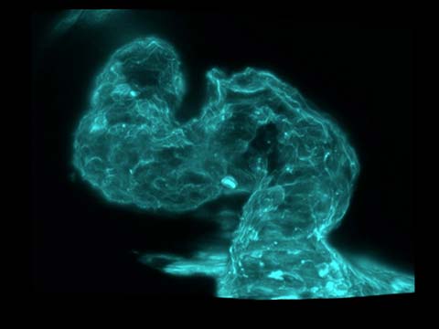
Dr. Andrew Moore captured this stunning video of neurons in an embryonic rat using a confocal microscope and fluorescent actin (Lifeact-EFGP) and is part of a larger experiment that set out to observe actin dynamics during neurite outgrowth. This movie was a combination of efforts which began with the transient transfection of a fluorescent actin sensor in to the neuron. Often the efficiency of this process is quite low so the next efforts required being able to optimize the microscope system to visualize the expression shown. In the end, it was all worth it, since this was the first time Dr. Moore had witnessed Neurite outgrowth. And of course, winning 4th Place in the Nikon Small World in Motion competition was an added bonus!
2020 Small World in Motion Competition
4th Place
Fluorescent actin (Lifeact-EGFP) expressed in an embryonic rat hippocampal neuron
Dr. Andrew Moore Dr. Pedro Guedes-Dias
- Affiliation
- Howard Hughes Medical Institute (HHMI)
Janelia Research Campus
Ashburn, Virginia, USA
- Technique
- Confocal
- Magnification
- 100X (Objective Lens Magnification)
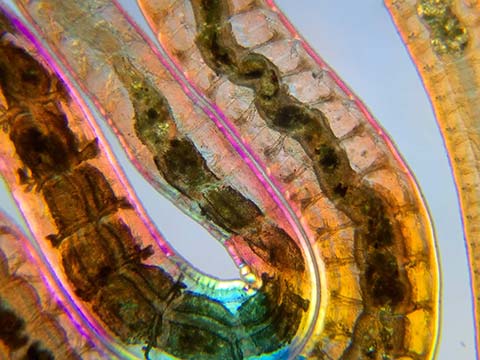
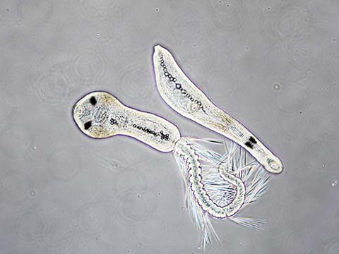
3rd Place
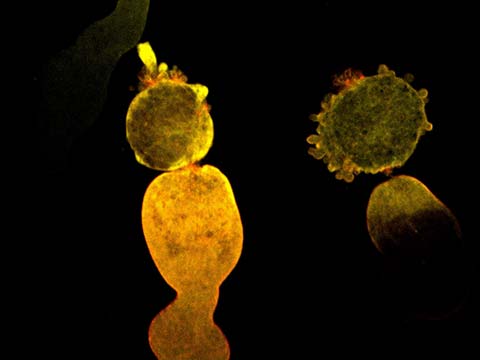
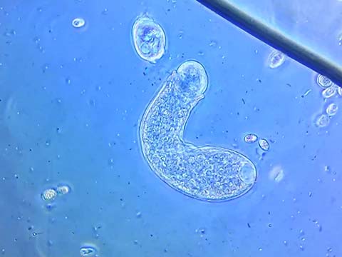
A blackworm (Lumbriculus variegatus) displaying peristaltic movements
Martin Kaae Kristiansen
- Affiliation
- My Microscopic World
Aalborg, Nordjylland, Denmark
- Technique
- Polarized Light
- Magnification
- 4X (Objective Lens Magnification)
This video of a blackworm, found in a pond water sample shows peristaltic (wavelike muscular contraction) movement inside the worm. Using a combination of optical staining and polarized light, it allowed Martin Kaae Kristiansen the ability to not only see inside the creature, but to observe and record its movements without hurting it. He finds this type of study important because it allows people to learn more about these tiny subjects and helps refine the lighting and staining techniques used in this and many other types of research.
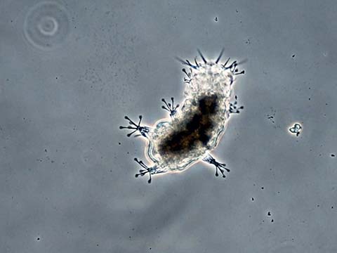
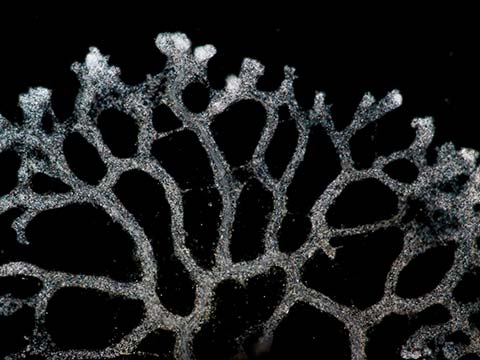
5th Place
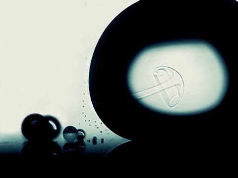
Crystallization of a callus removal solution
Wojtek Plonka
- Location
- Krakow, Malopolskie, Poland
- Technique
- Polarized Light
- Magnification
- 6.3X (Objective Lens Magnification)
The 5th place movie shows crystals from an over-the-counter callus remover growing as moisture is removed from the sample. Although Mr. Plonka’s main work involves computer simulations in chemistry (primarily for the purpose of drug design), he enjoys capturing stunning images of crystallization through the microscope as a sideline. Callus remover, which is a combination of lactic acid and salicylic acid, is a particularly popular sample due to its ability to crystalize into beautiful structures. It does require practice and talent to capture crystallization at this quality. From an equipment perspective, however, callus removal solution crystals can be observed with a standard high school science laboratory microscope.
Top 20
Honorable Mentions
Judges
Dr. Dylan Burnette
Assistant Professor of Cell and Developmental Biology Vanderbilt University

Burnette has been using high resolution microscopy to study cells for over 20 years. His laboratory at Vanderbilt University focuses on how cells grow and divide. He is interested in how these processes contribute to the function of heart muscle. He trained as a graduate student with Dr. Paul Forscher at Yale University and as a post-doctoral fellow with Dr. Jennifer Lippincott-Schwartz at the National Institutes of Health. Dr. Burnette has placed in the Nikon Small World competition eleven times.
Samantha Clark
Photo Editor National Geographic
Clark works on stories about science and the environment. She previously worked on NPR’s photo team and at Pier 24 Photography. Before working in visuals, she was a reporter and radio producer based in the Bay Area of San Francisco.
Sean Greene
Graphics and Data Journalist The Los Angeles Times
Greene covers science, the environment and medicine. He started working for The Los Angeles Times in 2014 and specializes in combining the powers of visual storytelling and the internet to tell meaningful and memorable stories. He’s reported on native oysters, bugs and frog tongues, and helped develop projects such as an interactive map of the Milky Way, a tracker of coronavirus cases in California and a data analysis of the dialogue in the Star Wars movies.
Dr. Christophe Leterrier
Group Leader Institute of Neurophysiopathology at CNRS and Aix-Marseille University
An engineer by training, Dr. Leterrier turned to cell biology and neurobiology for his Ph.D. He studies how neurons are organized at the cellular level and how they differentiate, then build and maintain their incredibly complex arborization. Since 2017, he has led the NeuroCyto lab in Marseille where he applies advanced microscopy techniques to directly observe molecular assemblies at the nanoscale inside neurons.
Ariel Waldman
Chair of the External Council NASA’s Innovative Advanced Concepts Program
Waldman led an expedition to Antarctica to film microscopic life under the ice. She is the co-author of a National Academy of Sciences report on the future of human spaceflight and the author of the book What’s It Like in Space?: Stories from Astronauts Who’ve Been There. Waldman is the global director of Science Hack Day, a National Geographic Explorer, a member of the San Francisco Microscopical Society, and received an honoree from the Obama White House as a Champion of Change in citizen science.