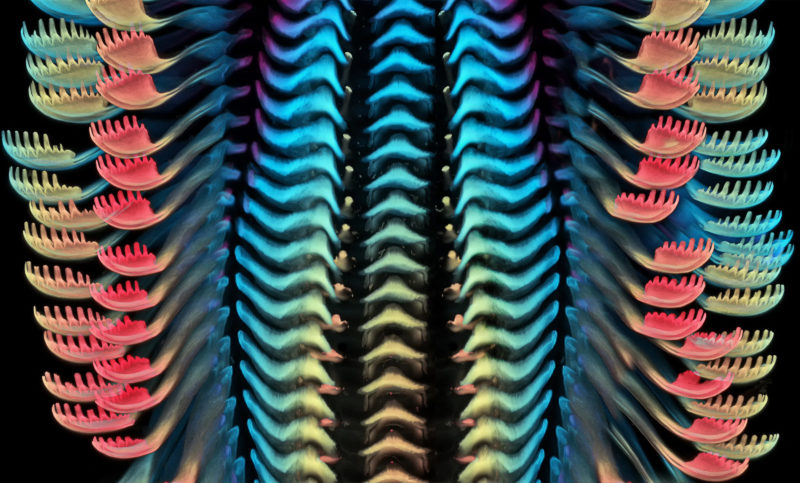
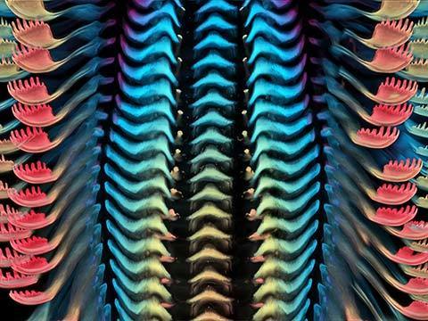
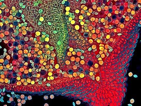
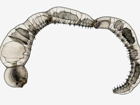
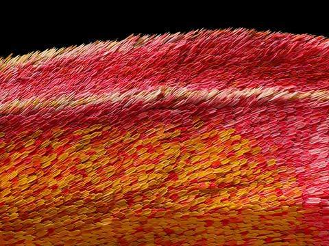
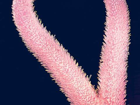
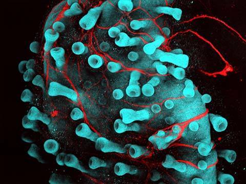
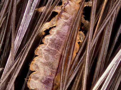
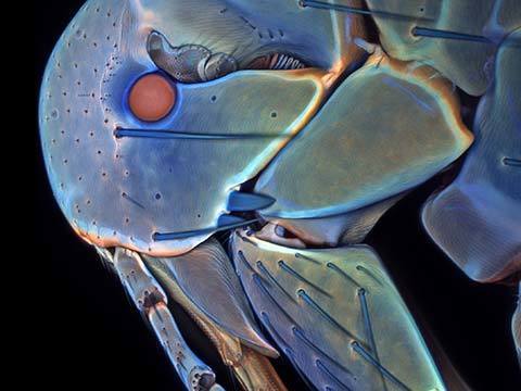
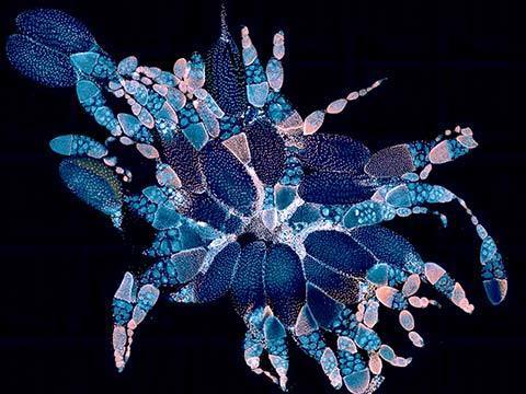
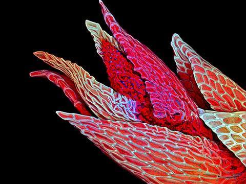
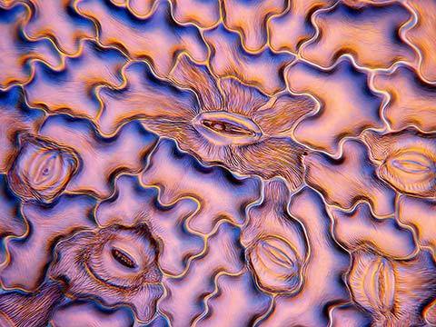
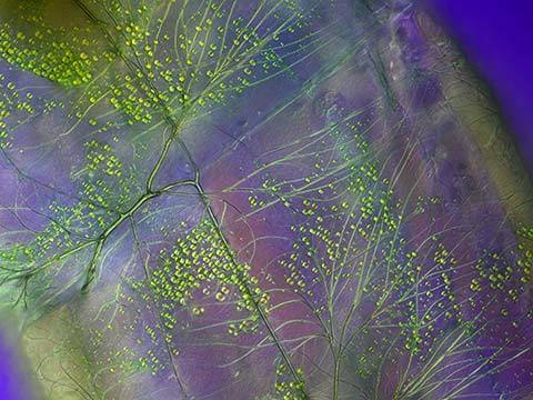
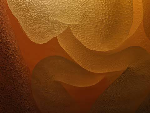
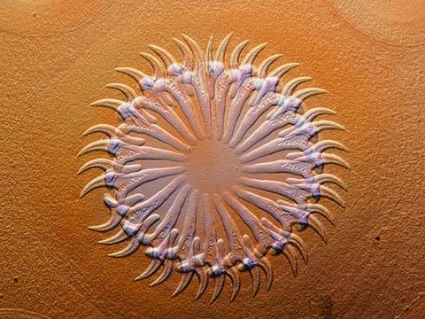
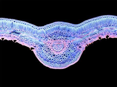
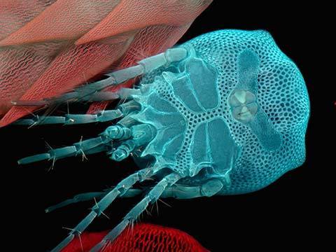
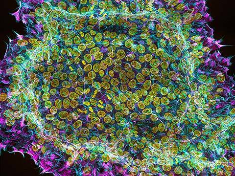
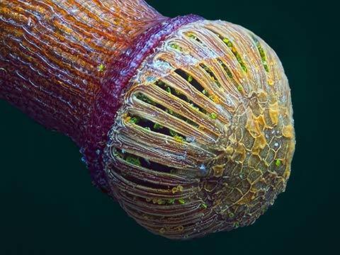
A depth color coded projection of a snail’s tongue (Radula) in Confocal, this image illustrates the beauty and complexity of natural forms even in something as seemingly simple as the tongue of a snail. The snail organ was frozen, stained with a chitin binding dye, mounted and captured using Confocal Z stacking.
2020 Photomicrography Competition
3rd Place
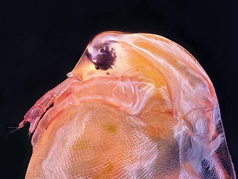
Tongue (radula) of a freshwater snail
Dr. Igor Robert Siwanowicz
- Affiliation
- Howard Hughes Medical Institute (HHMI)
Janelia Research Campus
Ashburn, Virginia, USA
- Technique
- Confocal
- Magnification
- 40X (Objective Lens Magnification)
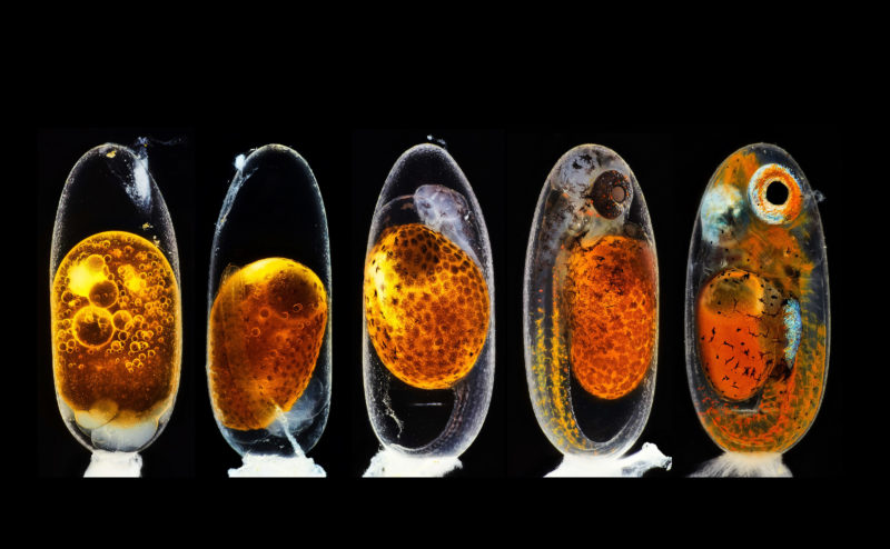
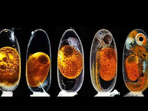
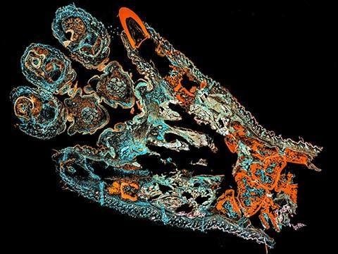
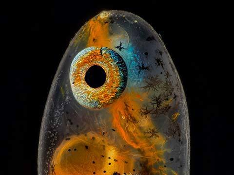
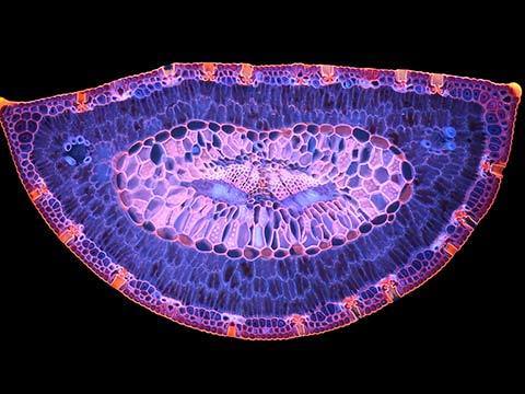
2nd Place
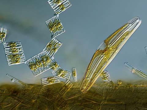
Embryonic development of a clownfish (Amphiprion percula) on days 1, 3 (morning and evening), 5, and 9
Daniel Knop
- Affiliation
- Natur und Tier-Verlag NTV
Oberzent-Airlenbach, Hessen, Germany
- Technique
- Image Stacking
- Magnification
- 10X (Objective Lens Magnification)
Daniel Knop's image of the embryonic development of a clownfish (Amphiprion percula) on days 1, 3 (morning and evening), 5, and 9, was created using image stacking. It shows the development, from hours after fertilization (even with a pack of sperm cells being visible on top of the egg), until hours before hatching. The primary challenge was to create sharp focus stacking pictures while the embryo was alive and moving.
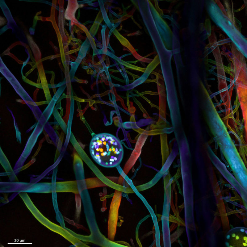
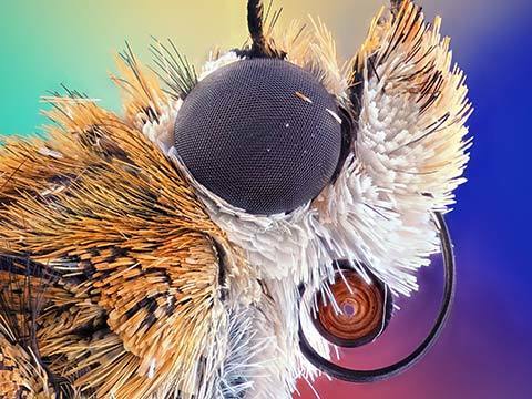
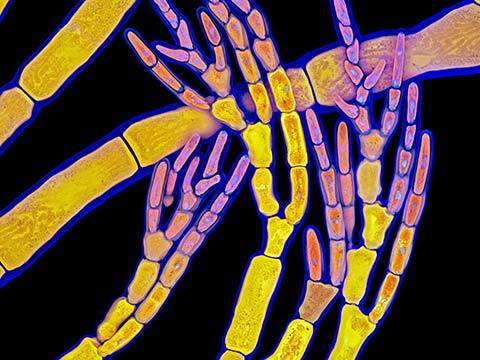
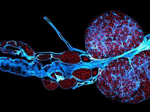
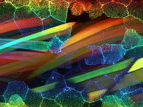
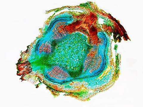
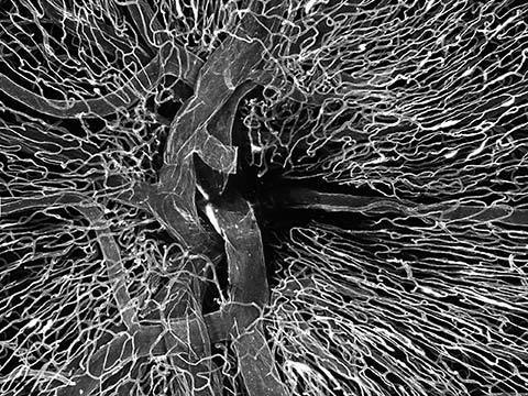
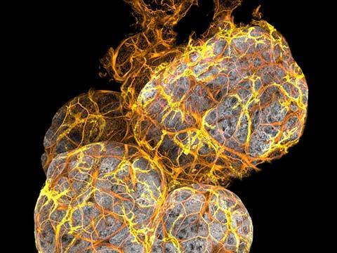
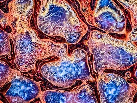
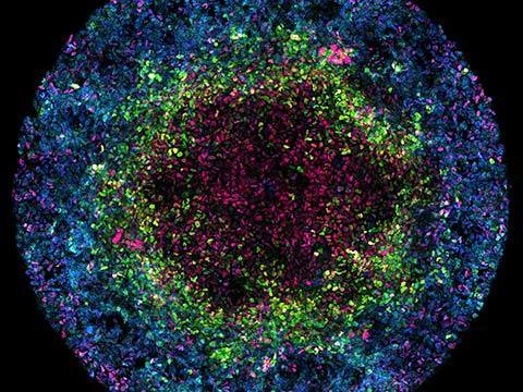
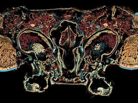
4th Place
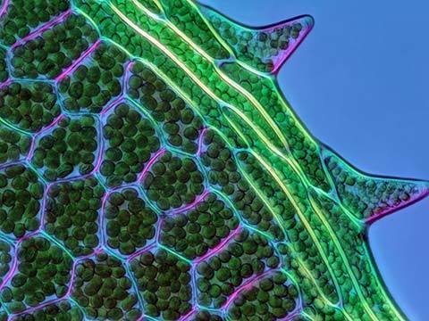
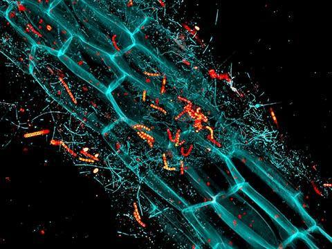
Multi-nucleate spores and hyphae of a soil fungus (arbuscular mycorrhizal fungus)
Dr. Vasileios Kokkoris Dr. Franck Stefani, Dr. Nicolas Corradi
- Affiliation
- Agriculture and Agri-Food Canada, University of Ottawa
Ottawa, Ontario, Canada
- Technique
- Confocal
- Magnification
- 63X (Objective Lens Magnification)
Dr. Vasileios Kokkoris captured this photo while researching fungal and plant ecology and the symbiotic relations between the two. It is the most commonly held notion that a cell contains a single nucleus within its structure, however the arbuscular mycorrhizal fungal cell carries multiple nuclei (shown) that can, in mature spores, reach hundreds or even thousands. This research is part of an ongoing study to understand the relationship between soil fungus and plants for agriculture. It was taken with a confocal microscope in order to visualize the nuclei as well as the hyphal and spore walls simultaneously.
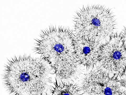
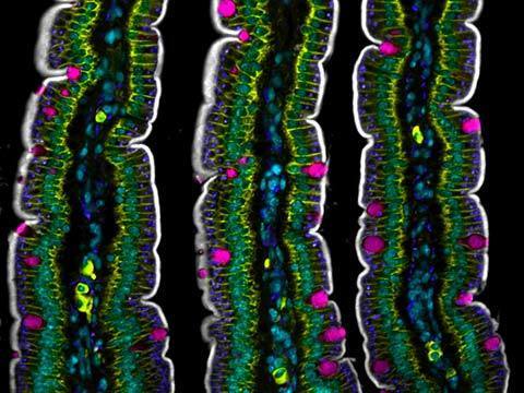
Top 20
Honorable Mentions
Images of Distinction
Judges
Dr. Dylan Burnette
Assistant Professor of Cell and Developmental Biology Vanderbilt University

Burnette has been using high resolution microscopy to study cells for over 20 years. His laboratory at Vanderbilt University focuses on how cells grow and divide. He is interested in how these processes contribute to the function of heart muscle. He trained as a graduate student with Dr. Paul Forscher at Yale University and as a post-doctoral fellow with Dr. Jennifer Lippincott-Schwartz at the National Institutes of Health. Dr. Burnette has placed in the Nikon Small World competition eleven times.
Samantha Clark
Photo Editor National Geographic
Clark works on stories about science and the environment. She previously worked on NPR’s photo team and at Pier 24 Photography. Before working in visuals, she was a reporter and radio producer based in the Bay Area of San Francisco.
Sean Greene
Graphics and Data Journalist The Los Angeles Times
Greene covers science, the environment and medicine. He started working for The Los Angeles Times in 2014 and specializes in combining the powers of visual storytelling and the internet to tell meaningful and memorable stories. He’s reported on native oysters, bugs and frog tongues, and helped develop projects such as an interactive map of the Milky Way, a tracker of coronavirus cases in California and a data analysis of the dialogue in the Star Wars movies.
Dr. Christophe Leterrier
Group Leader Institute of Neurophysiopathology at CNRS and Aix-Marseille University
An engineer by training, Dr. Leterrier turned to cell biology and neurobiology for his Ph.D. He studies how neurons are organized at the cellular level and how they differentiate, then build and maintain their incredibly complex arborization. Since 2017, he has led the NeuroCyto lab in Marseille where he applies advanced microscopy techniques to directly observe molecular assemblies at the nanoscale inside neurons.
Ariel Waldman
Chair of the External Council NASA’s Innovative Advanced Concepts Program
Waldman led an expedition to Antarctica to film microscopic life under the ice. She is the co-author of a National Academy of Sciences report on the future of human spaceflight and the author of the book What’s It Like in Space?: Stories from Astronauts Who’ve Been There. Waldman is the global director of Science Hack Day, a National Geographic Explorer, a member of the San Francisco Microscopical Society, and received an honoree from the Obama White House as a Champion of Change in citizen science.