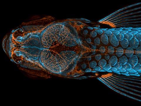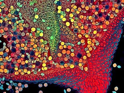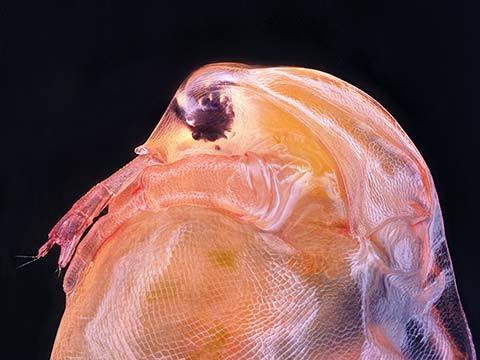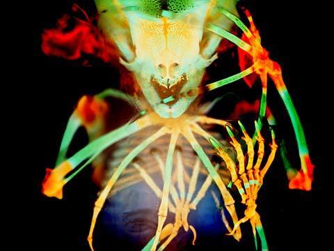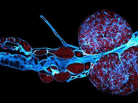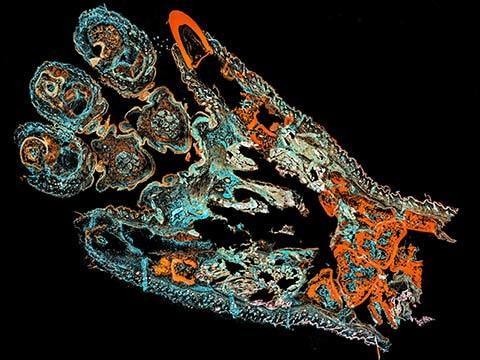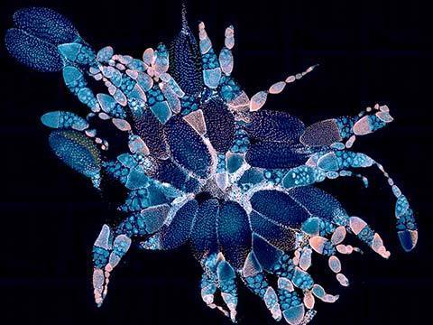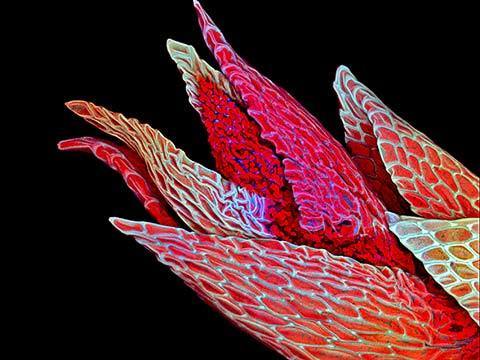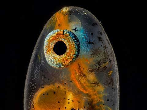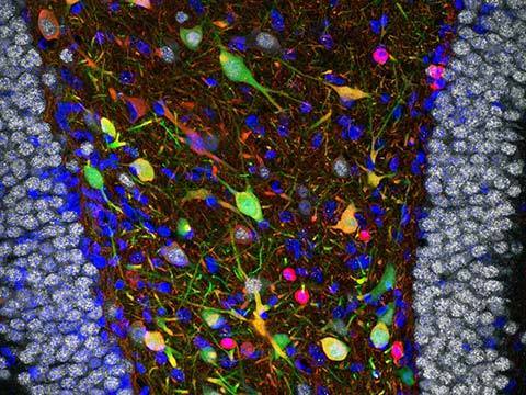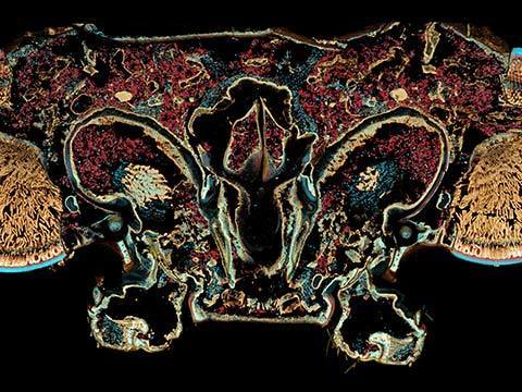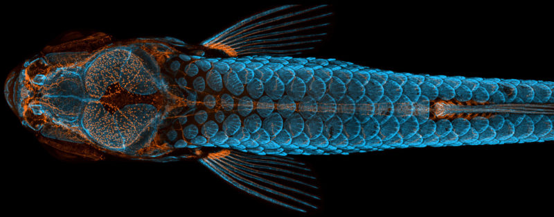
The stunning image that captured first place in the 2020 Nikon Small World Photomicrography Competition is a dorsal view of the head of a fish with fluorescently “tagged” skeleton, scales (blue) and lymphatic system (orange), taken using confocal microscopy and image-stacking.
This image is particularly significant because it was taken as part of an imaging effort that helped Castranova’s team make a groundbreaking discovery - zebrafish have lymphatic vessels inside their skull that were previously thought to occur only in mammals. Their occurrence in fish, a much easier subject to raise, experiment with, and photograph, could expedite and revolutionize research related to treatments for diseases that occur in the human brain, including cancer and Alzheimer’s.
Castranova stitched together more than 350 individual images to create this single stunning visual. The image was acquired using a spinning disk confocal, merging together maximum intensity projections of three separate image Z stacks to generate the final reconstructed image.
“The image is beautiful, but also shows how powerful the zebrafish can be as a model for the development of lymphatic vessels,” Castranova said, “Until now, we thought this type of lymphatic system only occurred in mammals. By studying them now, the scientific community can expedite a range of research and clinical innovations – everything from drug trials to cancer treatments. This is because fish are so much easier to raise and image than mammals.”