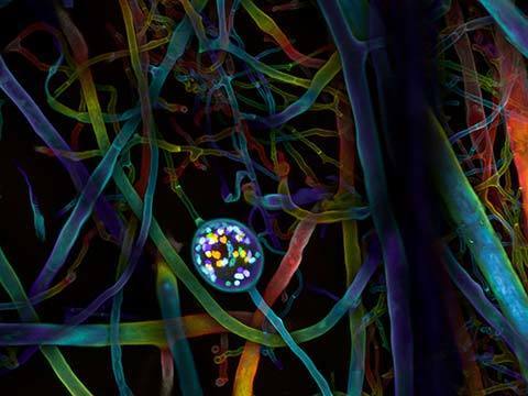
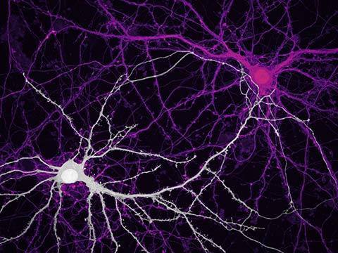
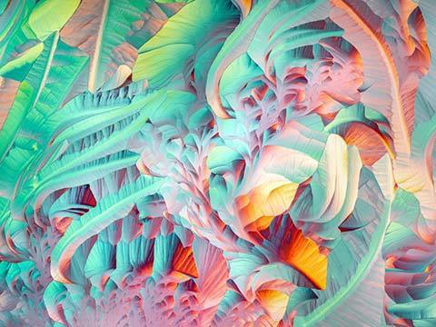
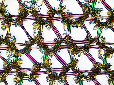
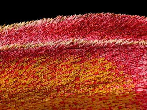
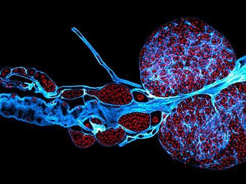
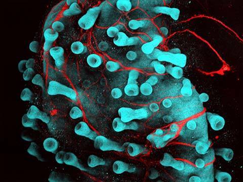
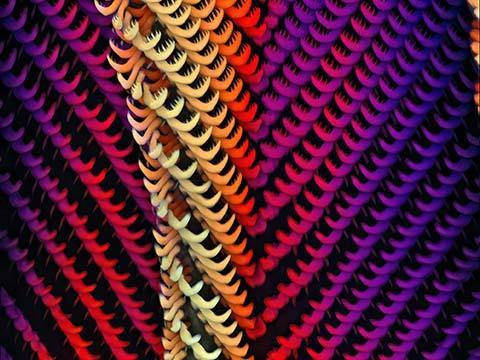
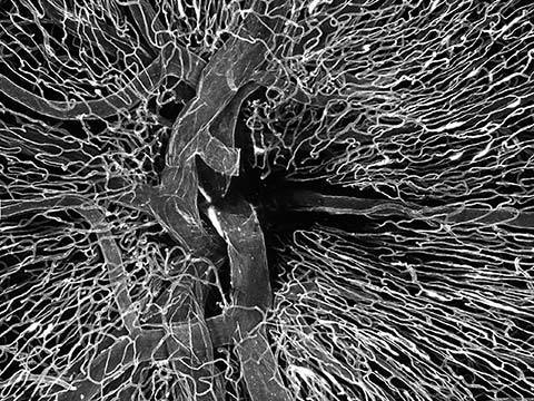
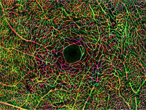
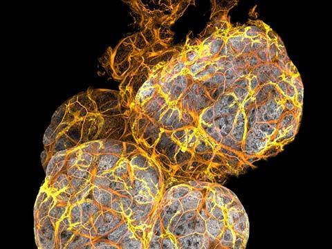
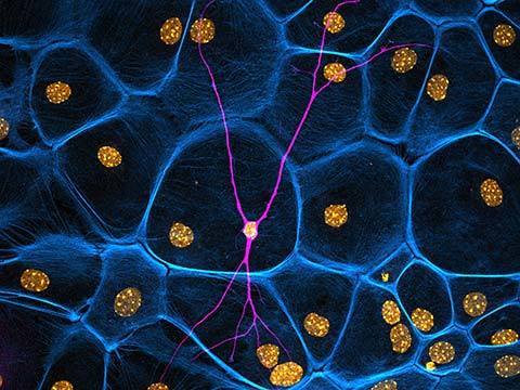
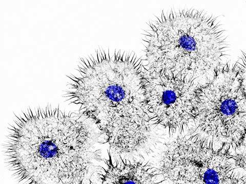
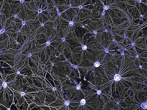
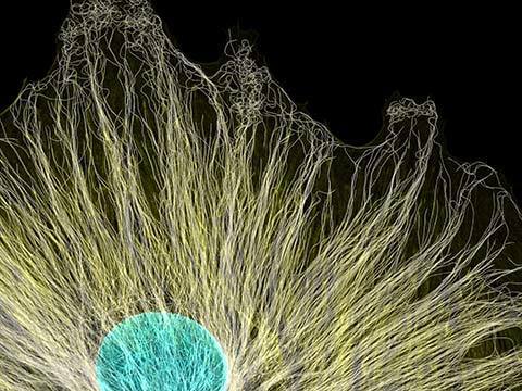
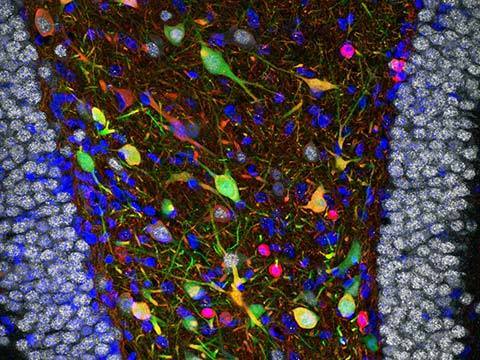
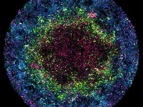
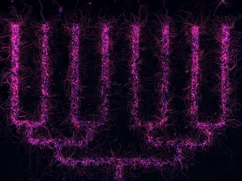
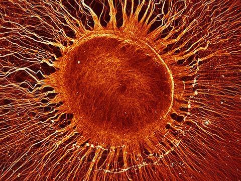
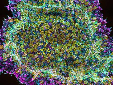
This is the second of three winning images from Jason Kirk in this year’s competition (see 7th place and his Image of Distinction). A professional microscopist and director of the Optical Imaging and Vital Microscopy Core facility at Baylor College of Medicine, Mr. Kirk took this image of neurons cultured from hippocampi of newly born mice. Note the level of detail and vast number of connections (dendritic spines) between these cells in this newly born specimen. This imaging is used in research of how certain proteins affect the development of dendritic spines.
2020 Photomicrography Competition
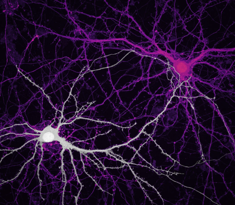
9th Place
Connections between hippocampal neurons (brain cells)
Jason Kirk Quynh Nguyen
- Affiliation
- Baylor College of Medicine
Optical Imaging & Vital Microscopy Core
Houston, Texas, USA
- Technique
- Confocal
- Magnification
- 63X (Objective Lens Magnification)
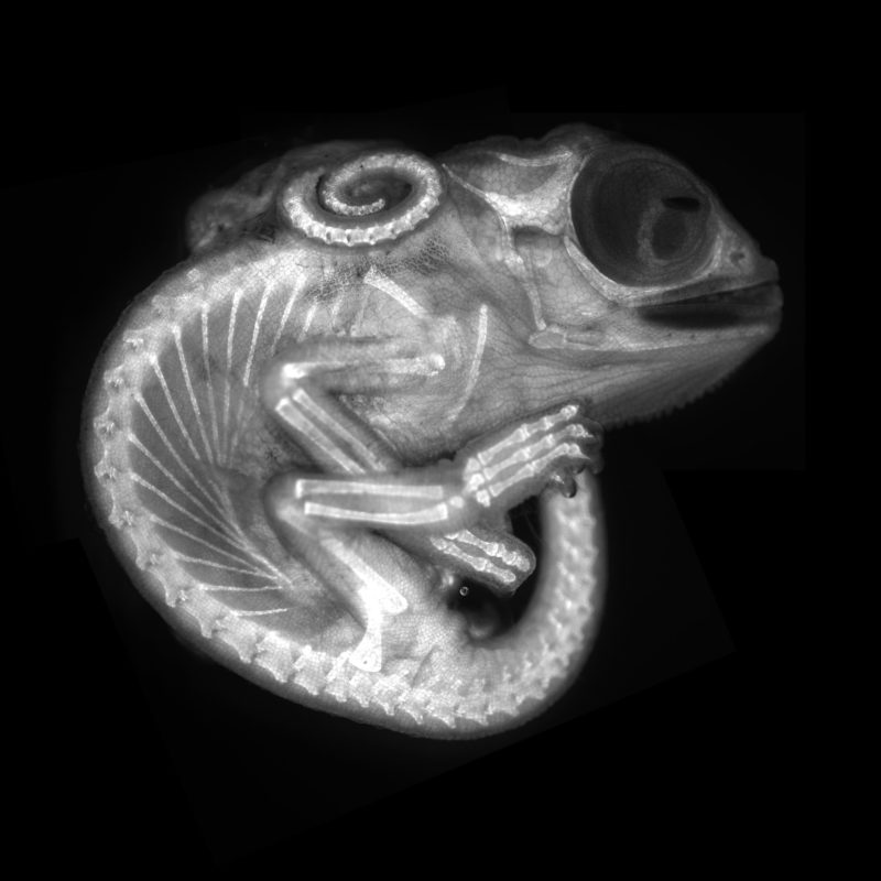
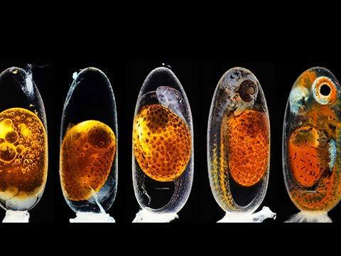
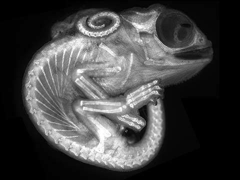
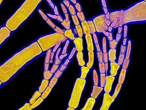
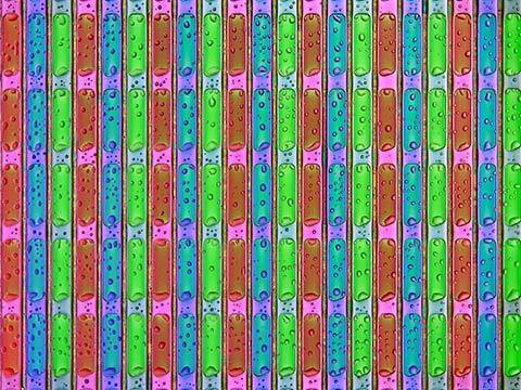
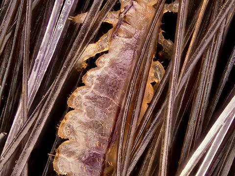
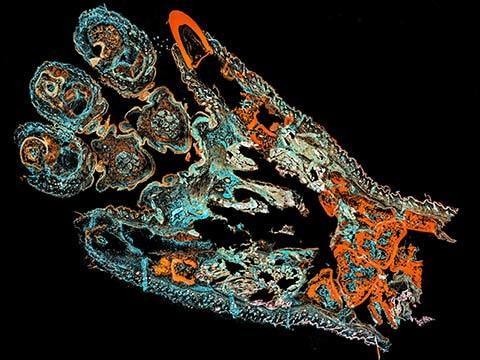
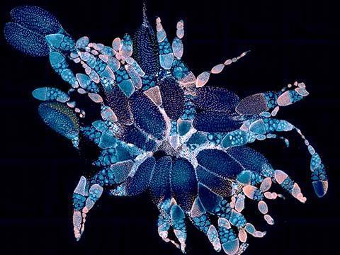
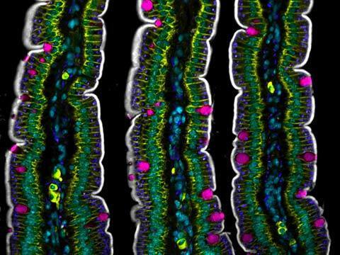
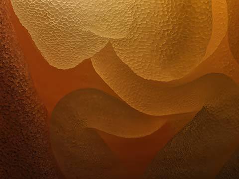
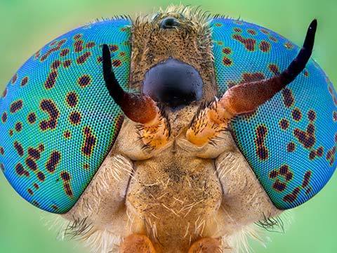
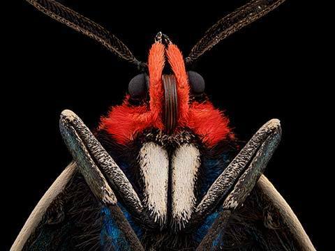
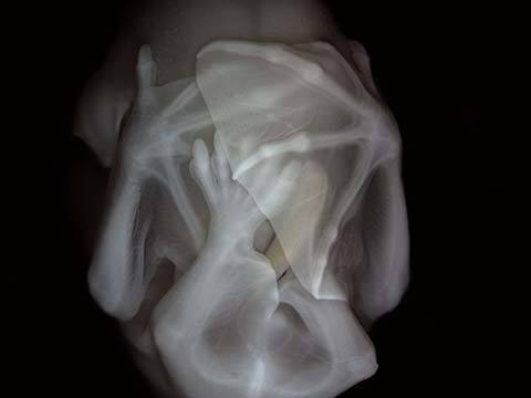
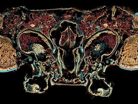
8th Place
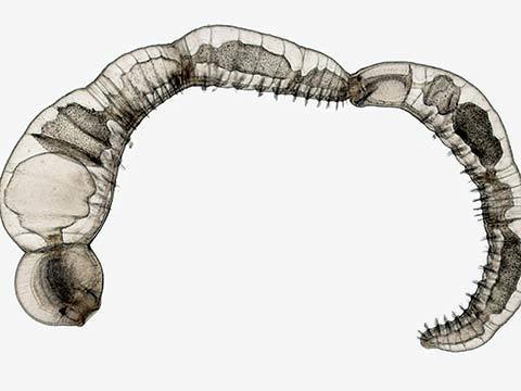
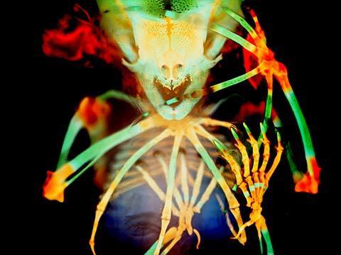
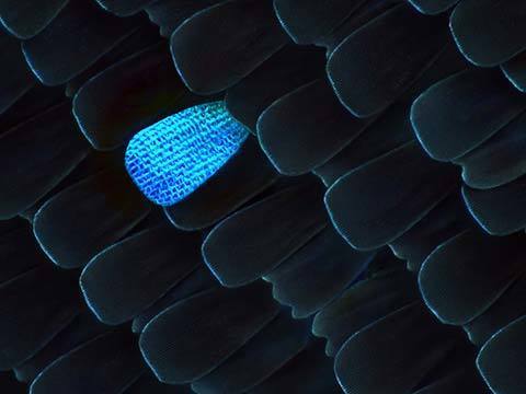
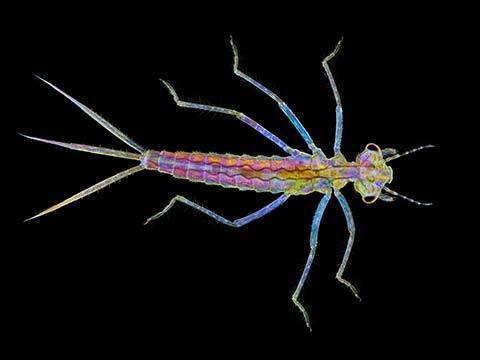
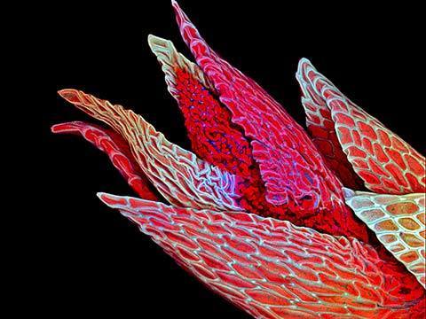
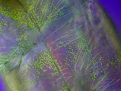
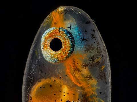
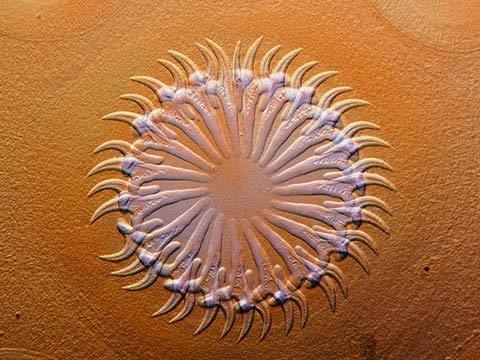
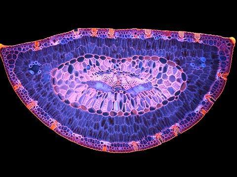
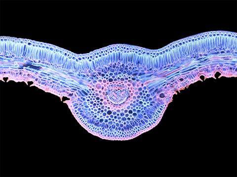
Chameleon embryo (autofluorescence)
Dr. Allan Carrillo-Baltodano David Salamanca
- Affiliation
- Queen Mary University of London
School of Biological and Chemical Sciences
London, United Kingdom
- Technique
- Fluorescence
- Magnification
- 10X (Objective Lens Magnification)
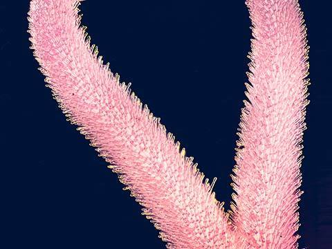
This image of a chameleon embryo, taken during the Embryology course at the Marine Biological Laboratory in Woods Hole, Massachusetts, highlights the aesthetics and structure of these invertebrates. It was two images with the subject submerged in liquid. Great care was necessary to avoid refraction from the liquid.
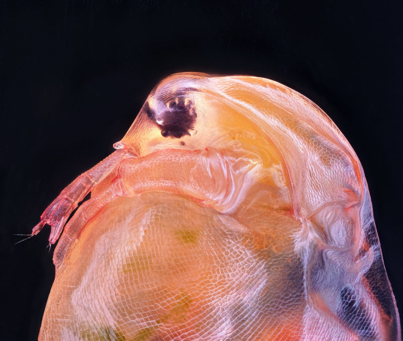
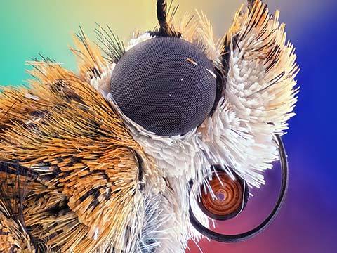
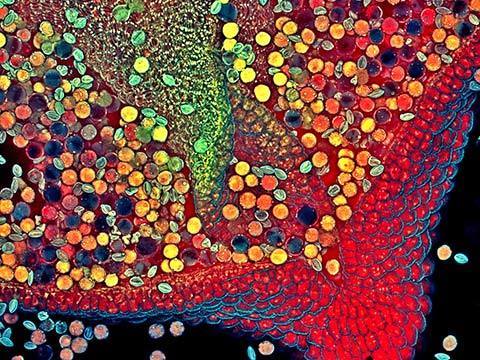
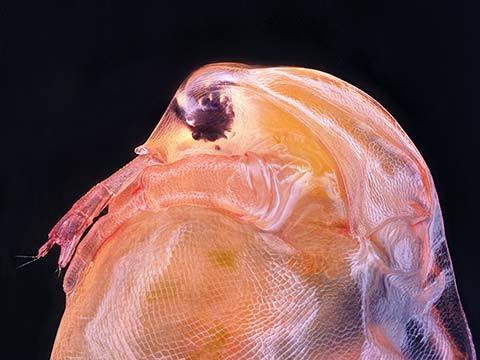
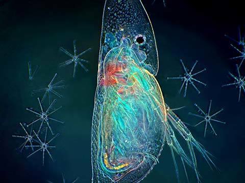
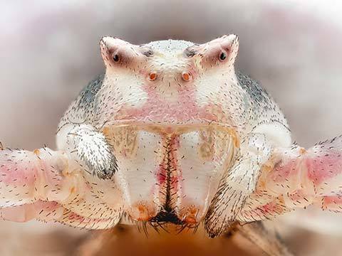
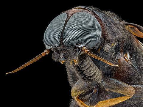
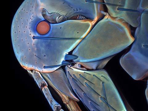
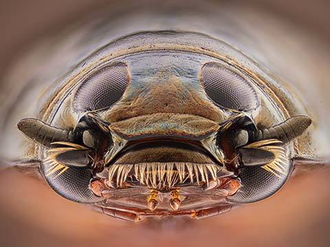
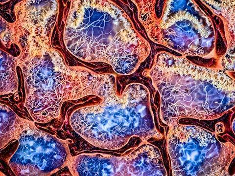
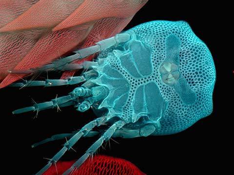
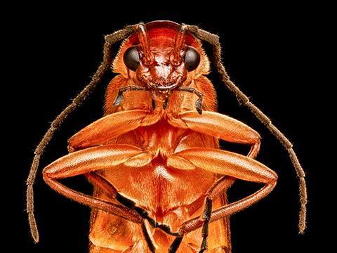
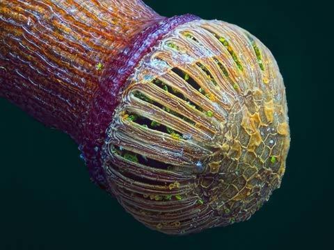
10th Place
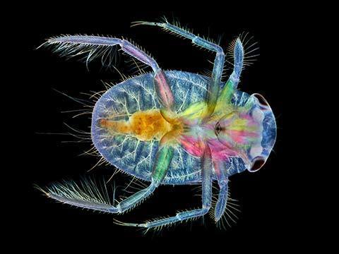
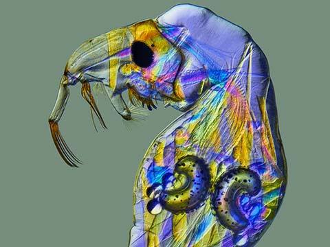
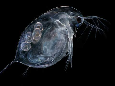
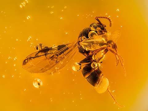
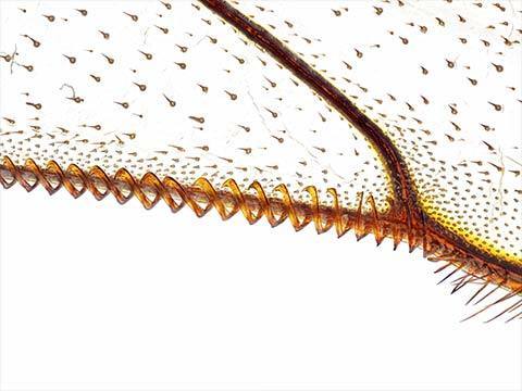
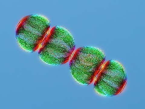
Daphnia magna (Phyllopoda)
Ahmad Fauzan
- Technique
- Image Stacking
- Magnification
- 10X (Objective Lens Magnification)
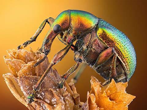
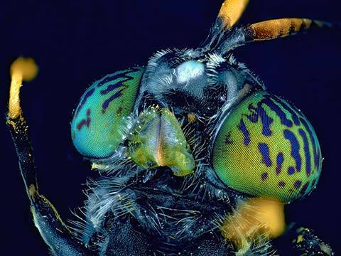
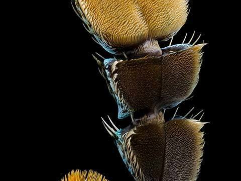
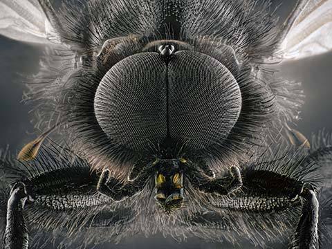
Mr. Fauzan, a multiple winner in 2020 (see 5th place), is an engineer by trade and a microscopist by hobby. This image is of Daphnia magna, which is a small planktonic crustacean and popular scientific study subject. The image was taken to illustrate the details of the eyes and antenna area. Focus and clarity was achieved using image stacking techniques.
Top 20
Honorable Mentions
Images of Distinction
Judges
Dr. Dylan Burnette
Assistant Professor of Cell and Developmental Biology Vanderbilt University

Burnette has been using high resolution microscopy to study cells for over 20 years. His laboratory at Vanderbilt University focuses on how cells grow and divide. He is interested in how these processes contribute to the function of heart muscle. He trained as a graduate student with Dr. Paul Forscher at Yale University and as a post-doctoral fellow with Dr. Jennifer Lippincott-Schwartz at the National Institutes of Health. Dr. Burnette has placed in the Nikon Small World competition eleven times.
Samantha Clark
Photo Editor National Geographic
Clark works on stories about science and the environment. She previously worked on NPR’s photo team and at Pier 24 Photography. Before working in visuals, she was a reporter and radio producer based in the Bay Area of San Francisco.
Sean Greene
Graphics and Data Journalist The Los Angeles Times
Greene covers science, the environment and medicine. He started working for The Los Angeles Times in 2014 and specializes in combining the powers of visual storytelling and the internet to tell meaningful and memorable stories. He’s reported on native oysters, bugs and frog tongues, and helped develop projects such as an interactive map of the Milky Way, a tracker of coronavirus cases in California and a data analysis of the dialogue in the Star Wars movies.
Dr. Christophe Leterrier
Group Leader Institute of Neurophysiopathology at CNRS and Aix-Marseille University
An engineer by training, Dr. Leterrier turned to cell biology and neurobiology for his Ph.D. He studies how neurons are organized at the cellular level and how they differentiate, then build and maintain their incredibly complex arborization. Since 2017, he has led the NeuroCyto lab in Marseille where he applies advanced microscopy techniques to directly observe molecular assemblies at the nanoscale inside neurons.
Ariel Waldman
Chair of the External Council NASA’s Innovative Advanced Concepts Program
Waldman led an expedition to Antarctica to film microscopic life under the ice. She is the co-author of a National Academy of Sciences report on the future of human spaceflight and the author of the book What’s It Like in Space?: Stories from Astronauts Who’ve Been There. Waldman is the global director of Science Hack Day, a National Geographic Explorer, a member of the San Francisco Microscopical Society, and received an honoree from the Obama White House as a Champion of Change in citizen science.