2019 Photomicrography Competition

Vitamin C

Karl Deckart

Location
Eckental, Bavaria, Germany
Technique
Brightfield, Polarized Light
Magnification
4x (Objective Lens Magnification)

In Their Own Words

A Q&A with Nikon Small World winner Karl Deckart.

What is the subject matter of your winning image and why did you choose this image?

The subject of this image is vitamin C. I chose to submit it because of its beauty.

What are the special techniques and/or challenges faced in creating this photomicrograph?

Polarized light.

How long have you been taking photographs through a microscope? What first sparked your interest in photomicrography?

33 years. Macrophotography led me to photomicrography.

Do you tend to focus your microscopy toward a specific subject matter or theme? If so, why?

I like to photograph anything that is eye-catching.

Housefly compound eye pattern

Dr. Razvan Cornel Constantin

Location
Bucharest, Romania
Technique
Focus Stacking, Reflected Light
Magnification
50x (Objective Lens Magnification)

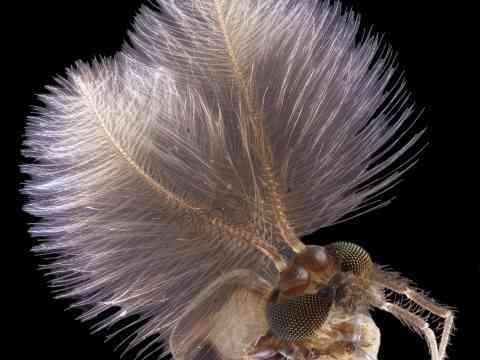
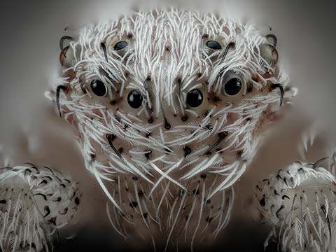
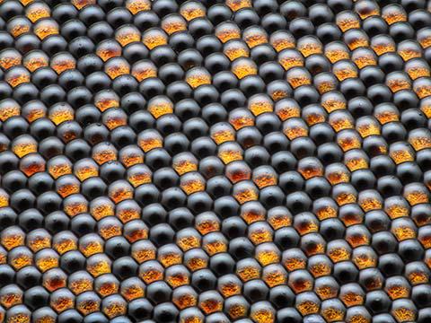
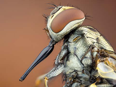
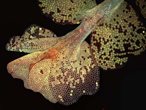
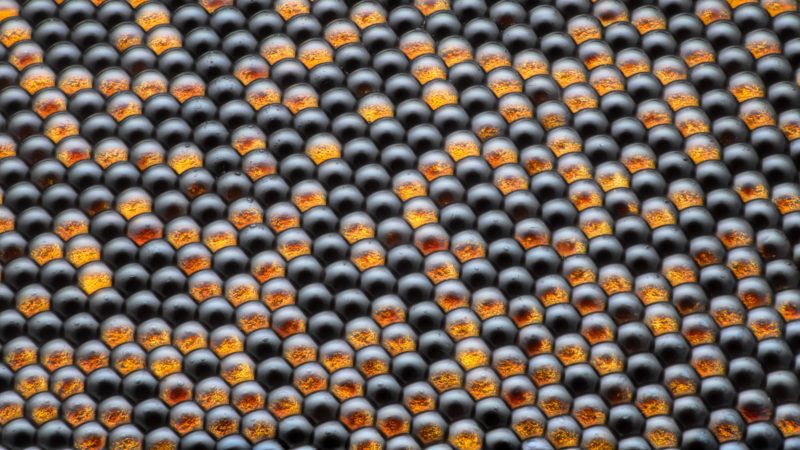
Flies look at the world in quite a different way than we do. Their eyes are made up of thousands of individual visual receptors called ommatidia, each of which is a functioning eye in itself. Therefore, a fly’s vision is comparable to a mosaic, with thousands of tiny images that converge together to represent one large visual image. The more ommatidia a compound eye contains, the clearer the image it creates.

A fly’s eyes are immobile, but their position and spherical shape give the fly an almost 360-degree view of its surroundings. Fly eyes have no pupils and cannot control how much light enters the eye or focus the images. Flies are also short-sighted — with a visible range of a few yards, and have limited color vision (for example, they don’t discern between yellow and white).

On the other hand, a fly’s vision is especially good at picking up form and movement. Because a fly can easily see motion but not necessarily what the moving object is, they are quick to flee, even if it is harmless.

In Their Own Words

A Q&A with Nikon Small World winner Dr. Razvan Cornel Constantin.

What is the subject matter of your winning image and why did you choose this image?

It is a closeup of a housefly decaying eye. The image doesn’t just show the structure of a compound eye but also what happens when the eye dries and the individual “cells” start to change color. It’s always a challenge to shoot at high magnification, and I thought this is a result worth sharing. The pattern is also very photogenic.

What are the special techniques and/or challenges faced in creating this photomicrograph?

For this picture I used focus stacking, which is challenging at high magnification because of the vibration of the camera and the rest of the equipment. At 50:1 the working distance is small for reflected light, so getting enough light onto the subject is always a struggle. Also, getting it diffused in such a way that the individual lenses on the eye reflect it in a pleasing way without losing detail was tricky.

What is your primary line of work?

I make my living as an automotive engineer, but when I get home and pick up my camera, that’s when the job stops and the passion begins.

How long have you been taking photographs through a microscope? What first sparked your interest in photomicrography?

I’ve been using microscopes for almost four years, gradually increasing the magnification as I got more experienced. I’ve always had a passion for wildlife, especially insects. As soon as I could afford it, I got a camera and macro lens. While shooting macro you always crave for more magnification and that’s why I got into photomicrography.

Do you tend to focus your microscopy toward a specific subject matter or theme? If so, why?

I can’t say that I have a specific subject, I find that almost any subject has at least a few interesting poses when put under a microscope at high magnification. As long as you can’t see it with the naked eye you always get that wow factor.

Cristobalite crystal suspended in its quartz mineral host

Billie Hughes

Affiliation
Lotus Gemology
Bangkok, Thailand
Technique
Darkfield
Magnification
40x (Objective Lens Magnification)

The mushroom floating in this photo is actually a crystal included in a piece of mineral.

Quartz is one of the most common minerals on earth and a constituent of many rocks. The crystal in the photo is cristobalite, a mineral composed of the same elements as quartz but with a different crystal structure.

Cristobalite is common in volcanic rocks and gets its name from Cerro San Cristobal, Mexico, where it is was first observed.

The major uses of cristobalite and quartz are in construction-related activities such as road building and as a cement additive. Other uses include the manufacturing of glass fibers, ceramics, rubber and coatings, and as an abrasive. Cristobalite is also used in dentistry as a component of impression materials as well as for making models of teeth.

In Their Own Words

A Q&A with Nikon Small World winner E. Billie Hughes.

What is the subject matter of your winning image and why did you choose this image?

A white, mushroom-shaped cristobalite crystal, which is suspended inside its quartz mineral host. I chose this image because I felt it is a good representation of how the field of gemology combines both art and science. While we study gems because of their beauty and rarity, we also use scientific techniques, particularly microscopy, to gain a deeper and more complete understanding of gems and minerals. When I saw this cristobalite crystal inside of quartz, my first thought was that it looks surprisingly similar to a mushroom. It is interesting to see how two such different creations of the natural world can so closely resemble each other.

What are the special techniques and/or challenges faced in creating this photomicrograph?

The first challenge in capturing this image was to align the mineral specimen at just the right angle so that the cristobalite inclusion would take on its mushroom shape. It was also important to get the lighting correct to illuminate the cristobalite crystal against its dark background. I used a combination of darkfield and carefully positioned diffuse fiber optic lights to achieve the desired effect for this image.

What is your primary line of work?

I am a gemologist at Lotus Gemology, a colored gemstone testing laboratory based in Bangkok, Thailand.

How long have you been taking photographs through a microscope? What first sparked your interest in photomicrography?

Approximately five years. I first became interested in photomicrography when I took gemology classes and started looking at gems through the microscope. When I looked at the internal world of gems, I felt like I was exploring a new frontier, and I wanted to share that with others through photomicrography.

Do you tend to focus your microscopy toward a specific subject matter or theme? If so, why?

I focus my microscopy on gemstone inclusions, which are the irregularities found inside gems. Microscopy can help gemologists learn a lot about gems, including whether they are natural or synthetic (man-made) and whether they have gone through any treatment, such as heat treatment. In some cases, it can even help us determine where in the world a gem was found.

Why did you enter the Nikon Small World Photomicrography competition? What do you think of the competition?

I entered this competition because I had seen previous years’ winners and was amazed and inspired by their stunning photomicrographs. I feel honored to be included among this talented group of people. Nikon has done a great job using this competition as a tool to promote microscopy and photomicrography, making what could otherwise be a niche field more accessible.