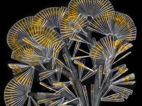
2019 Photomicrography Competition
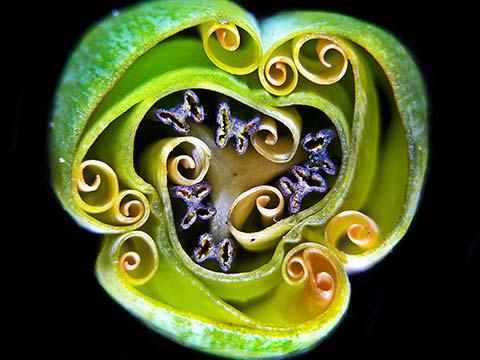
9th Place
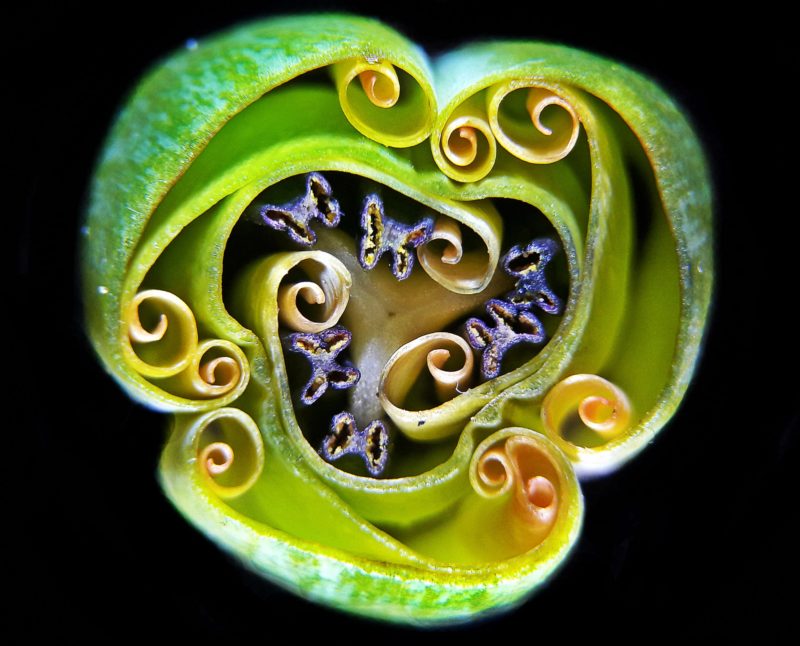
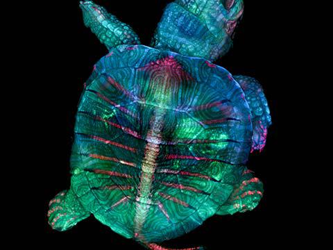
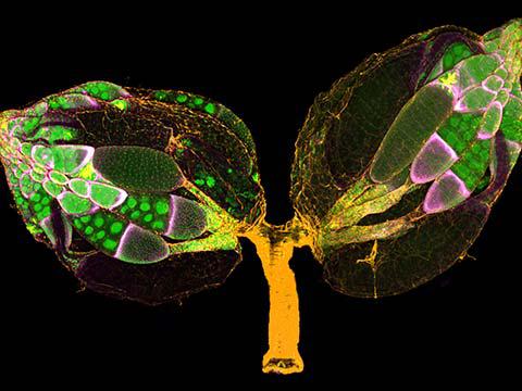
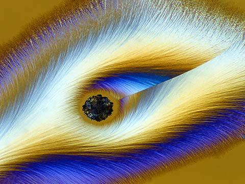
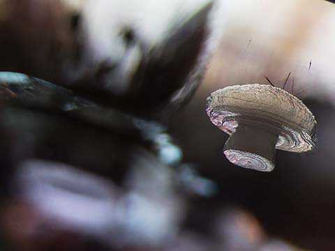
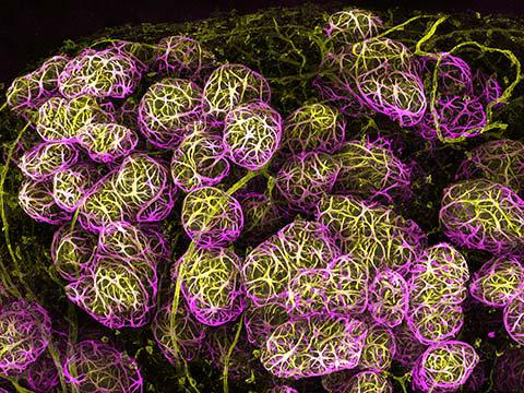
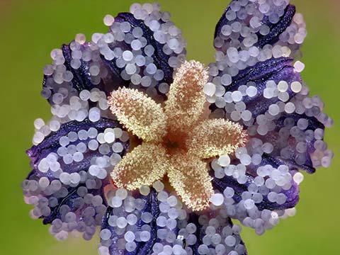
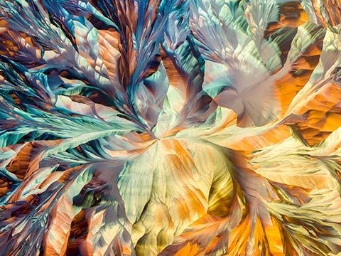
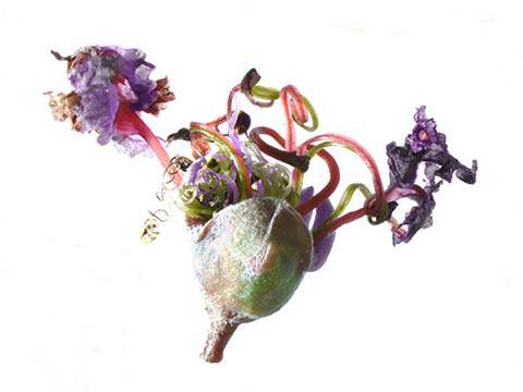
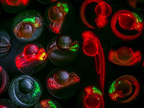
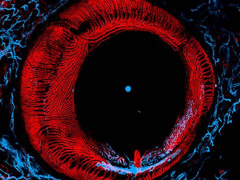
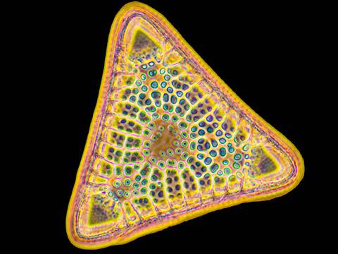
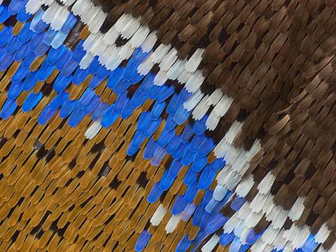
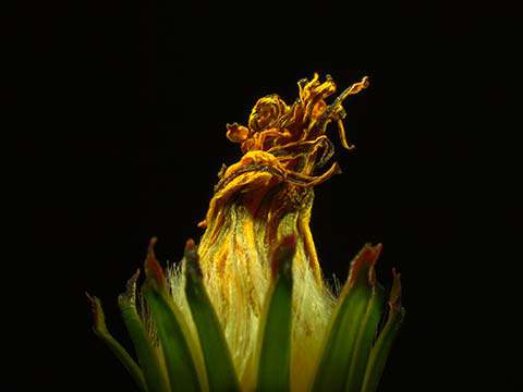
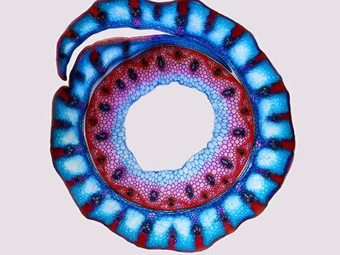
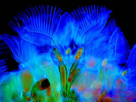
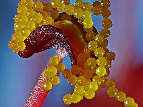
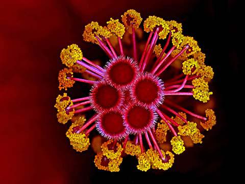
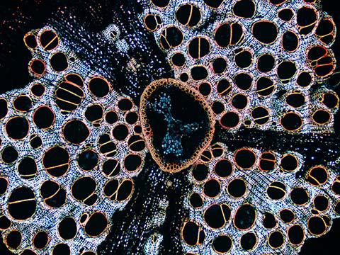
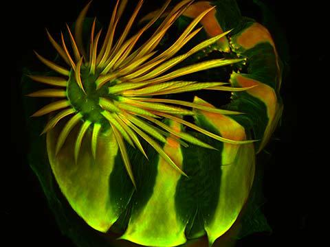
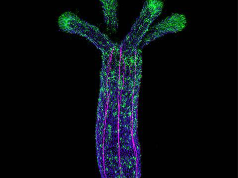
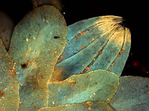
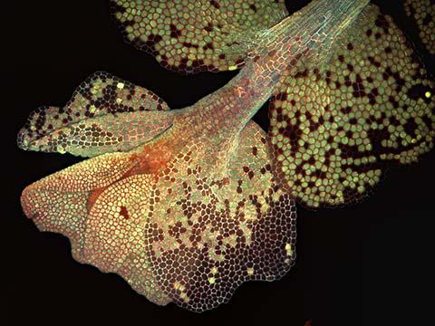
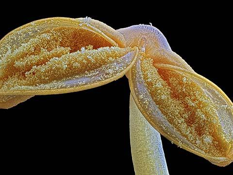
Tulip bud cross section
Andrei Savitsky
- Location
- Cherkassy, Ukraine
- Technique
- Reflected Light
- Magnification
- 1x (Objective Lens Magnification)
In Their Own Words
A Q&A with Nikon Small World winner Andrei Savitsky.
What is the subject matter of your winning image and why did you choose this image?
The internal structure of an undiscovered tulip bud. I submitted it because when I look at this photo I am fascinated by its symmetry of the spiral structures. We never see what is inside a bud, we always admire the flower, but I can show that a nondescript bud can be more beautiful than the flower itself.
What are the special techniques and/or challenges faced in creating this photomicrograph?
The problem is that the diameter of the bud is more than 1 cm, so even at the lowest magnification, it is impossible to photograph. I came up with the idea of using a phase telescope as a macro lens. I put the phase telescope in the microscope tube and removed the objective lens. And took the photo through the phase telescope.
What is your primary line of work?
I am an electrical engineer by education, but I dream that microphotography will become my main work.
How long have you been taking photographs through a microscope? What first sparked your interest in photomicrography?
I started in 2011, after a microscope demonstration in a school class. When I got the microscope, everything was a discovery for me, I wanted to show it to others to share my delight with them.
Do you tend to focus your microscopy toward a specific subject matter or theme? If so, why?
No. I try to capture all the topics I can.
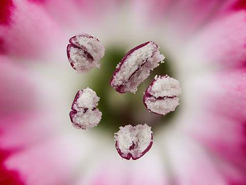
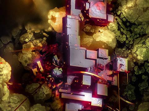
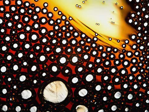
8th Place
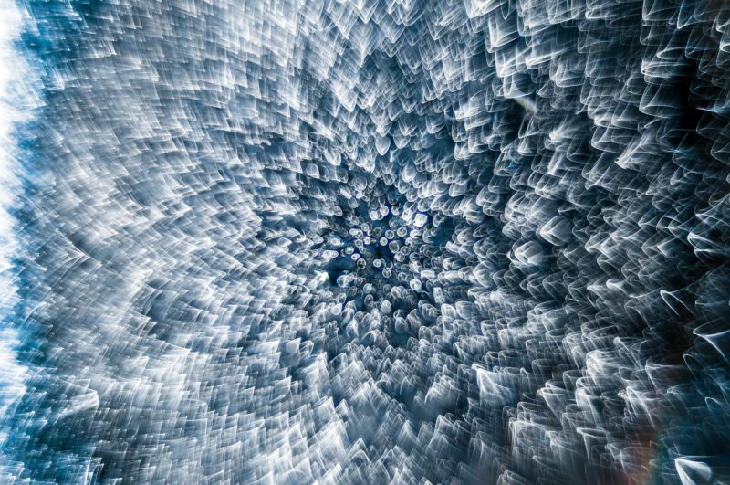
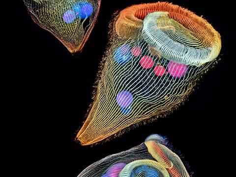
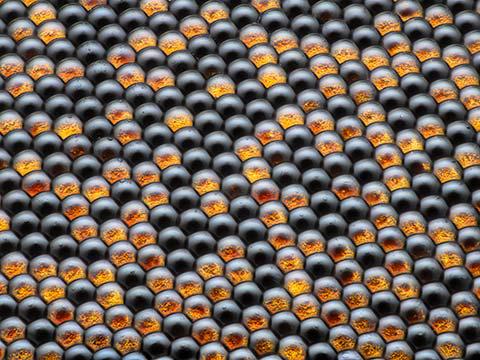
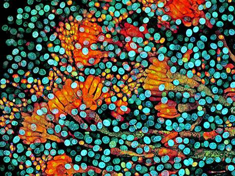
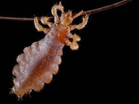
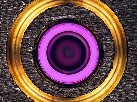
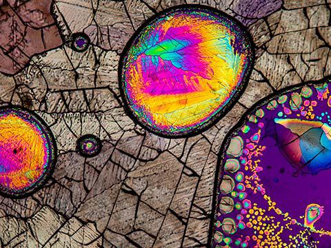
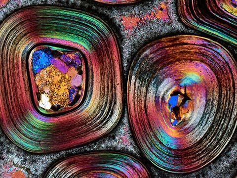
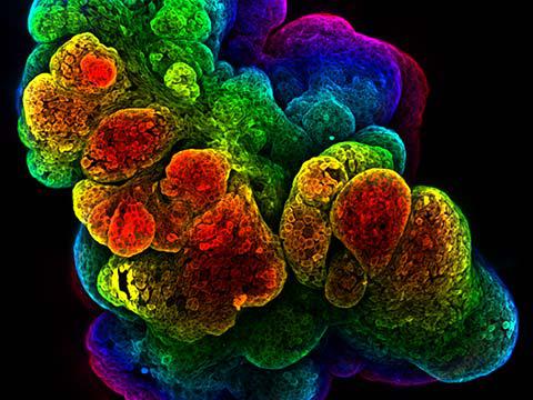
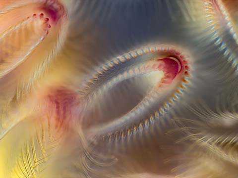
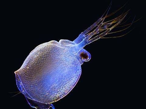
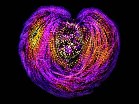
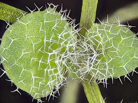
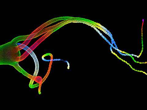
Frozen water droplet
Garzon Christian
- Location
- Quintin, Cotes-d’Armor, France
- Technique
- Incident Light
- Magnification
- 8x (Objective Lens Magnification)
In Their Own Words
A Q&A with Nikon Small World winner Garzon Christian.
What is the subject matter of your winning image and why did you choose this image?
A frozen water droplet that was on my car hood in sunny conditions. I chose this image because it surprised me after a test shoot, as it looked different from what my eyes saw. I like this picture for its beauty and the surprise of the final render. This reminded me of what happens in my job (I am a social educator) when I realize that something special and beautiful hides behind the first impression I get of a person.
What are the special techniques and/or challenges faced in creating this photomicrograph?
Sadly, I can’t describe the process because the final result of this picture is due to a mistake in my preparation. I did not respect the distance between my lens and my sensor, so it’s challenging to reproduce the same setting.
How long have you been taking photographs through a microscope? What first sparked your interest in photomicrography?
I have been practicing photography through a microscope in the past six months. I was amazed by the possibility to discover nature’s hidden beauty.
Do you tend to focus your microscopy on a specific subject matter or theme? If so, why?
I just like to have fun with the camera and the microscope, I like to get “inside” things.
Why did you enter the Nikon Small World Photomicrography competition? What do you think of the competition?
I entered the Nikon Small World competition to offer my point of view of nature. I like this competition because it’s open to everyone and we can find inspiration.
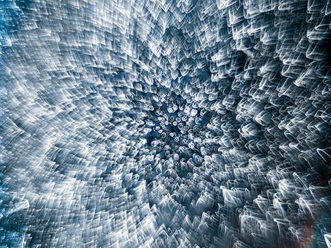
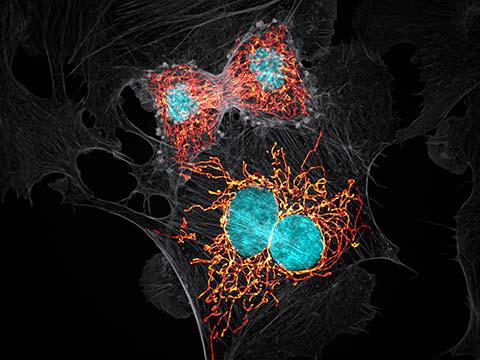
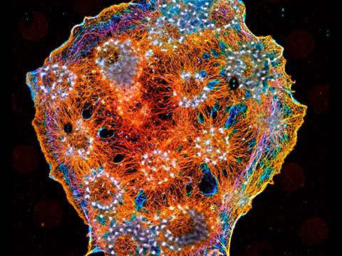
10th Place
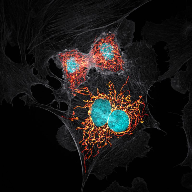
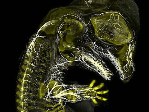
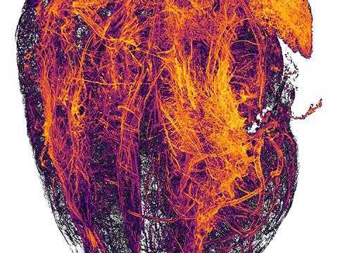
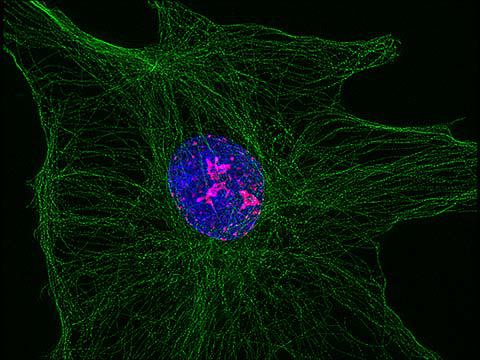
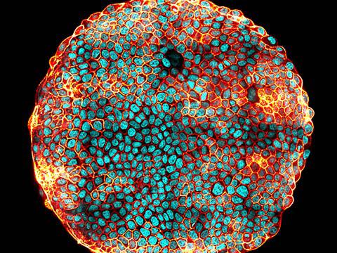
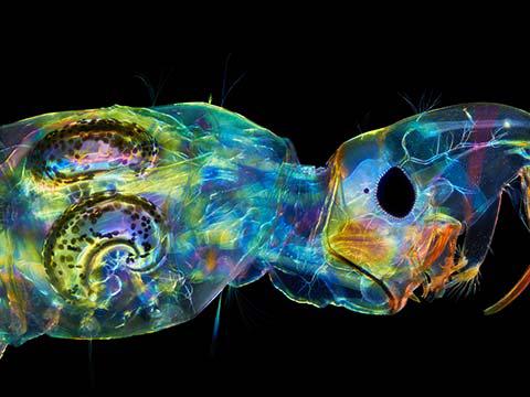
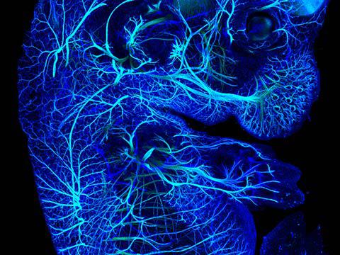
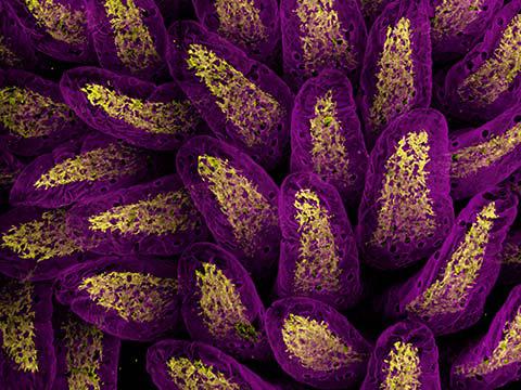
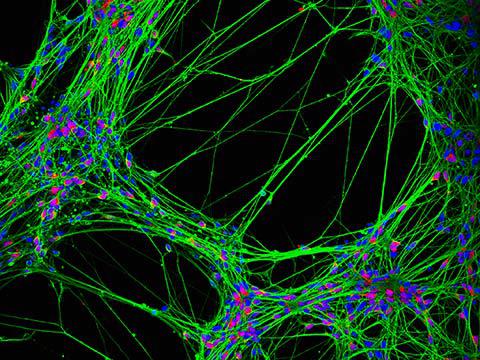
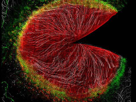
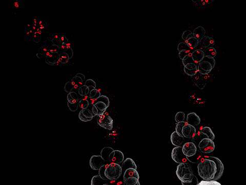
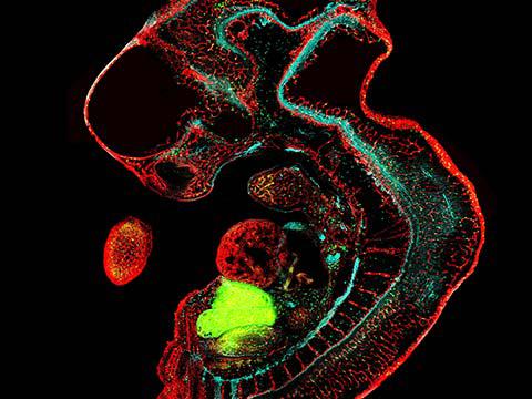
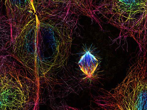
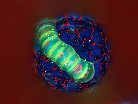
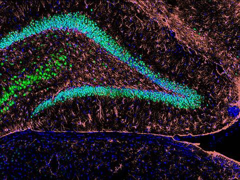
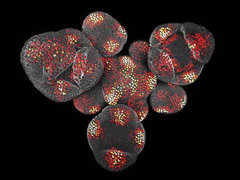
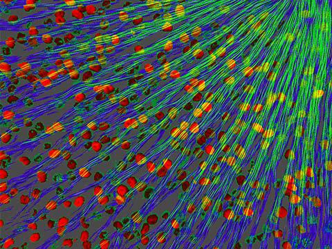
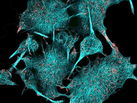
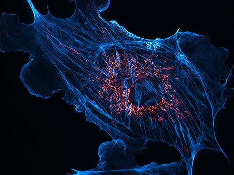
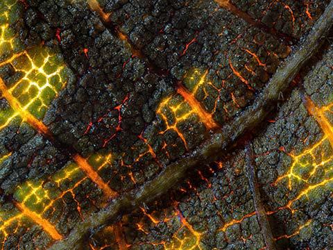
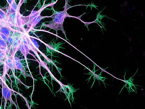
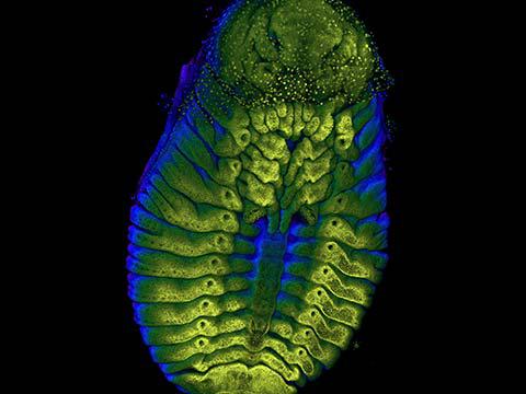
BPAE cells in telophase stage of mitosis
Jason Kirk
- Affiliation
- Baylor College of Medicine
Optical Imaging & Vital Microscopy Core
Houston, Texas, USA
- Technique
- Confocal with Enhanced Resolution
- Magnification
- 63x (Objective Lens Magnification)
In Their Own Words
A Q&A with Nikon Small World winner Jason Kirk.
What is the subject matter of your winning image and why did you choose this image?
Bovine Pulmonary Artery Endothelial cells with mitochondria labeled with Mitotracker (glow scale), F-Actin (grey) and DAPI (magenta) for DNA. It is one of the strongest compositions I have made with this cell line, which is a wonderful benchmark tool for microscopists. These cells are fascinating to image due to the endless variations in morphology and number of markers that can be used. I wanted to illustrate the idea that mitochondria are the power plants of the cell. Fire is synonymous with energy and the contrast of the monochrome ‘ashes’ of the cytoskeleton against the fiery mitochondria illustrates this nicely, I believe.
What are the special techniques and/or challenges faced in creating this photomicrograph?
This was a time-consuming image to get balanced correctly. Each of the three channels had to be imaged separately as well as processed independently to produce the final composition. To get everything in focus, a maximum intensity projection was done on each Z stack and the image was pseudo-colored in postproduction.
What is your primary line of work?
I direct a core facility for the Baylor College of Medicine that focuses on fluorescence imaging of large intact tissue models using 3-D optical sectioning tools.
How long have you been taking photographs through a microscope? What first sparked your interest in photomicrography?
More than 20 years. I had an undergraduate advisor who was passionate about imaging flatworms. He introduced me to electron and fluorescence microscopy. This was in the late 90s when digital imaging was just starting to be used but was very expensive. The first time I took a picture from a computer I was hooked.
Do you tend to focus your microscopy toward a specific subject matter or theme? If so, why?
I am a bit of a generalist. Our researchers are interested in a vast array of model systems and so, as a core facility director, I have broad access to many different subjects. Fluorescence has always interested me because, while it is a tool targeted at the molecular level, we can observe the labeling over large areas of intact tissue, which makes the imaging exciting and challenging.
Why did you enter the Nikon Small World Photomicrography competition? What do you think of the competition?
NSW is a fantastic showcase for a diverse community of photographers made up of scientists, hobbyists and artists, which blends traditional photography and science in a way that is not often seen in mainstream media. I think this competition reminds people that the world is so much bigger than what they can see with their eyes and I wanted to be a part of that.
Top 20

Honorable Mentions

Images of Distinction


Judges
Dr. Denisa Wagner
Edwin Cohn Professor of Pediatrics at Harvard Medical School and the Head of the Wagner Lab Harvard Medical School and Boston Children’s Hospital
Dr. Rita Strack
Senior Editor Nature Methods
Tom Hale
Staff Writer IFLScience
Ben Guarino
Science Reporter The Washington Post