2019 Photomicrography Competition

Cristobalite crystal suspended in its quartz mineral host

Billie Hughes

Affiliation
Lotus Gemology
Bangkok, Thailand
Technique
Darkfield
Magnification
40x (Objective Lens Magnification)

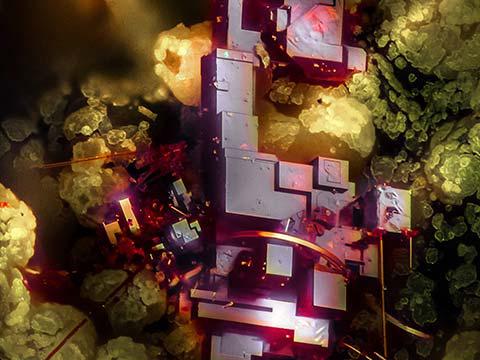
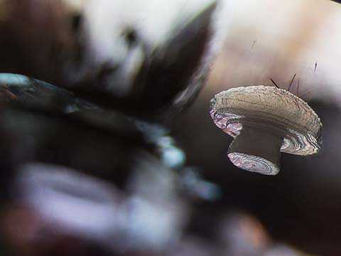
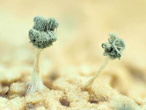
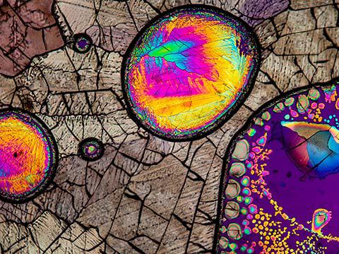
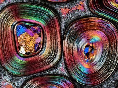
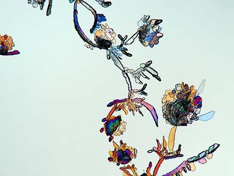
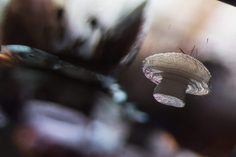
The mushroom floating in this photo is actually a crystal included in a piece of mineral.

Quartz is one of the most common minerals on earth and a constituent of many rocks. The crystal in the photo is cristobalite, a mineral composed of the same elements as quartz but with a different crystal structure.

Cristobalite is common in volcanic rocks and gets its name from Cerro San Cristobal, Mexico, where it is was first observed.

The major uses of cristobalite and quartz are in construction-related activities such as road building and as a cement additive. Other uses include the manufacturing of glass fibers, ceramics, rubber and coatings, and as an abrasive. Cristobalite is also used in dentistry as a component of impression materials as well as for making models of teeth.

In Their Own Words

A Q&A with Nikon Small World winner E. Billie Hughes.

What is the subject matter of your winning image and why did you choose this image?

A white, mushroom-shaped cristobalite crystal, which is suspended inside its quartz mineral host. I chose this image because I felt it is a good representation of how the field of gemology combines both art and science. While we study gems because of their beauty and rarity, we also use scientific techniques, particularly microscopy, to gain a deeper and more complete understanding of gems and minerals. When I saw this cristobalite crystal inside of quartz, my first thought was that it looks surprisingly similar to a mushroom. It is interesting to see how two such different creations of the natural world can so closely resemble each other.

What are the special techniques and/or challenges faced in creating this photomicrograph?

The first challenge in capturing this image was to align the mineral specimen at just the right angle so that the cristobalite inclusion would take on its mushroom shape. It was also important to get the lighting correct to illuminate the cristobalite crystal against its dark background. I used a combination of darkfield and carefully positioned diffuse fiber optic lights to achieve the desired effect for this image.

What is your primary line of work?

I am a gemologist at Lotus Gemology, a colored gemstone testing laboratory based in Bangkok, Thailand.

How long have you been taking photographs through a microscope? What first sparked your interest in photomicrography?

Approximately five years. I first became interested in photomicrography when I took gemology classes and started looking at gems through the microscope. When I looked at the internal world of gems, I felt like I was exploring a new frontier, and I wanted to share that with others through photomicrography.

Do you tend to focus your microscopy toward a specific subject matter or theme? If so, why?

I focus my microscopy on gemstone inclusions, which are the irregularities found inside gems. Microscopy can help gemologists learn a lot about gems, including whether they are natural or synthetic (man-made) and whether they have gone through any treatment, such as heat treatment. In some cases, it can even help us determine where in the world a gem was found.

Why did you enter the Nikon Small World Photomicrography competition? What do you think of the competition?

I entered this competition because I had seen previous years’ winners and was amazed and inspired by their stunning photomicrographs. I feel honored to be included among this talented group of people. Nikon has done a great job using this competition as a tool to promote microscopy and photomicrography, making what could otherwise be a niche field more accessible.

Vitamin C

Karl Deckart

Location
Eckental, Bavaria, Germany
Technique
Brightfield, Polarized Light
Magnification
4x (Objective Lens Magnification)

In Their Own Words

A Q&A with Nikon Small World winner Karl Deckart.

What is the subject matter of your winning image and why did you choose this image?

The subject of this image is vitamin C. I chose to submit it because of its beauty.

What are the special techniques and/or challenges faced in creating this photomicrograph?

Polarized light.

How long have you been taking photographs through a microscope? What first sparked your interest in photomicrography?

33 years. Macrophotography led me to photomicrography.

Do you tend to focus your microscopy toward a specific subject matter or theme? If so, why?

I like to photograph anything that is eye-catching.

Octopus bimaculoides embryo

Martyna Lukoseviciute Dr. Carrie Albertin

Affiliation
University of Oxford
Weatherall Institute of Molecular Medicine
Oxford, Oxfordshire, United Kingdom
Technique
Confocal, Image Stitching
Magnification
5x (Objective Lens Magnification)

In Their Own Words

A Q&A with Nikon Small World winner Martyna Lukoseviciute.

What is the subject matter of your winning image and why did you choose this image?

It’s an octopus embryo. Octopus is my favorite sea animal. I am fascinated by its intelligence, camouflage abilities and social interactions, but it is very different from mammals. Therefore, it is important to study its development, especially the nervous system development that can hold answers regarding the conserved and basic features of intelligent animals. This image is very beautiful and also educates people on the importance of understanding marine animal development.

What are the special techniques and/or challenges faced in creating this photomicrograph?

The biggest challenge was mounting the embryo onto the slide, because of its relatively big size. I needed to use a specialized slide with a little groove and build little walls around the sample to put the coverslip on top of it to avoid squashing the embryo. Also, because of its size, I needed to use a 5x objective and acquire multiple files to cover the whole area of the sample. This took a long time but was really worth it!

What is your primary line of work?

I am in my last year of my Ph.D. in developmental biology. I am researching neural crest specification and early differentiation controlled by epigenetic mechanisms in the zebrafish embryo.

How long have you been taking photographs through a microscope? What first sparked your interest in photomicrography?

I started taking photographs through a microscope in 2015 during my bachelor’s degree, when I was working on frog embryos. However, I have only started using high-resolution confocal microscopes during my DPhil at the University of Oxford for my work with zebrafish embryos. Seeing is believing! I was mesmerized by the beautiful structures hidden from our eyes and once I found a way to remove this barrier by using microscopes, I completely fell in love with it. I love sharing and engaging people outside the scientific community by showing my microscopy pictures of different species' embryos.

Do you tend to focus your microscopy toward a specific subject matter or theme? If so, why?

Usually, my microscopy is focused on embryo development. Nothing fascinates me more than unraveling the secrets of embryo development and comparing different species.

Why did you enter the Nikon Small World Photomicrography competition? What do you think of the competition?

I entered this competition because every year I am excited to see the winning images and learn something new about both the world and different techniques. I was excited to share my octopus image with people like me — excited to learn something new and admire the hidden beauty. As a scientist, it’s relevant to me to share images of such embryos, showing how important it is to study and compare the basic principles of embryo development. Hopefully, images like this can bring the attention back to basic science, marine biology and evolution.