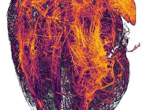
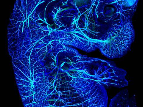
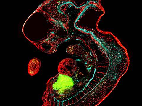
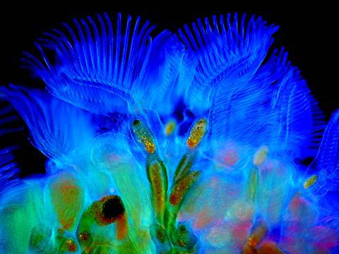
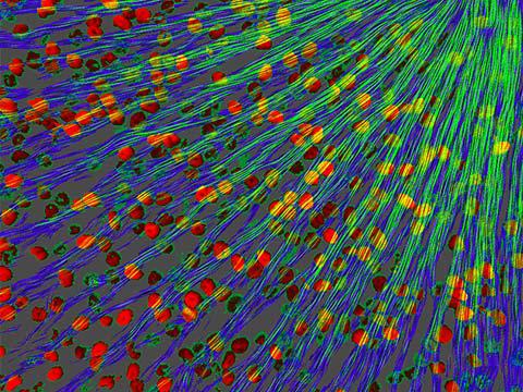
2019 Photomicrography Competition
20th Place
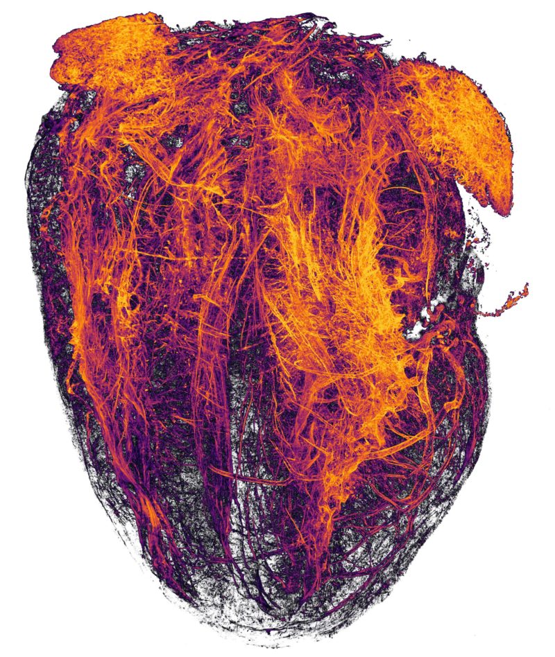
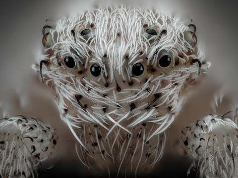
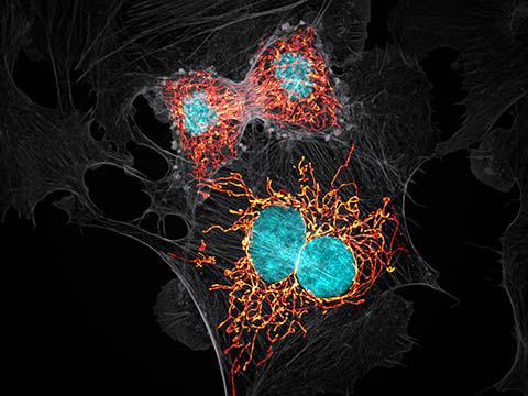
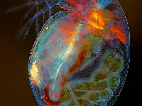
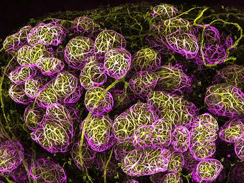
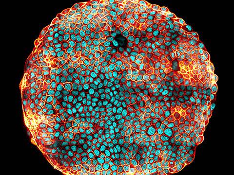
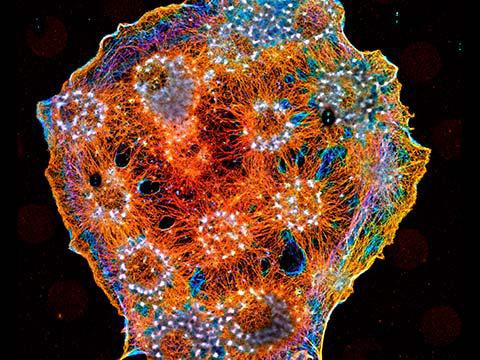
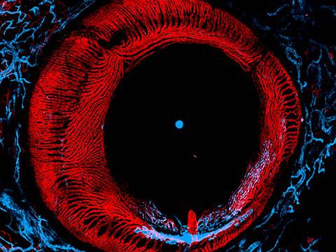
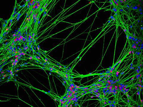
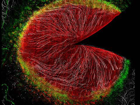
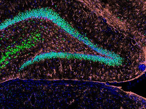
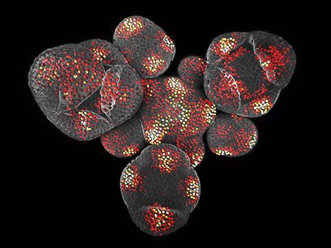
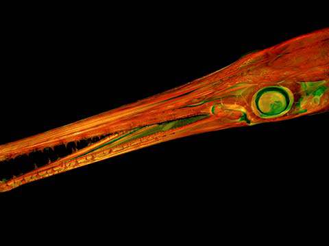
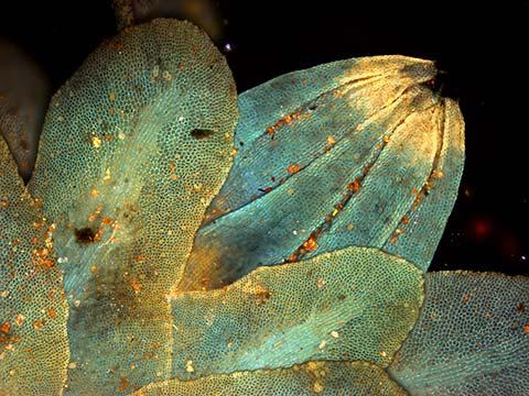
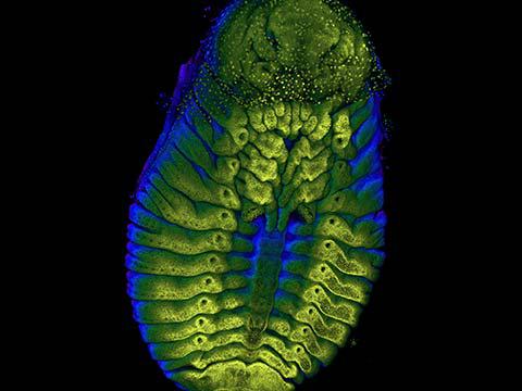
Blood vessels of a murine (mouse) heart following myocardial infarction (heart attack)
Dr. Simon Frederik Merz Dr. Lea Bornemann, Sebastian Korste
- Affiliation
- University Hospital Essen
Institute for Experimental Immunology & Imaging
Essen, Nordrhein-Westfalen, Germany
- Magnification
- 2x (Objective Lens Magnification)
In Their Own Words
A Q&A with Nikon Small World winner Simon Merz.
What is the subject matter of your winning image and why did you choose this image?
The blood vessel system (mainly arteries) of a whole murine heart after an induced heart attack. Raw data were algorithm-enhanced to achieve better contrast. I am working towards the introduction of this technique in routine clinical diagnostics and for some tests we obtain proof of principle of our protocols using animal tissues before we can work with human material. The protocol used to clear this sample was later developed into a technique to clear and analyze a metastatic human lymph node. This image is a great example of what can be done with light-sheet fluorescence 3-D microscopy in combination with tissue clearing, giving an overview of the complex organ structure. It shows a whole mouse heart with high resolution, providing an easy-to-grasp and new perspective on the body.
What are the special techniques and/or challenges faced in creating this photomicrograph?
The challenge here lies in the clearing process before imaging. The sample needs to be translucent, so light can pass through it without being scattered to a high degree. Therefore, we have to change the composition of the tissue to homogenize the refractive index of all components within the sample. This, in combination with fluorophore-labeled antibodies against blood vessels, allows an in-depth analysis of the entire specimen. However, the obtained information is so dense that showing a 2-D representation of the 3-D data stack of about 6 mm tissue depth would result in a chaotic image. Therefore, algorithms are developed to clear images of distracting information, enhancing structural details that are relevant and allowing to focus on specific structures.
What is your primary line of work?
I am a Ph.D. student in the field of medical biology. I work on the implementation of novel 3-D microscopy in routine cancer diagnostics, mainly in the field of melanoma.
How long have you been taking photographs through a microscope? What first sparked your interest in photomicrography?
Since 2012, during my bachelor studies in Heidelberg, Germany. I started with simple nuclear staining. Typically, to study what happens inside a cell, an organ or body, we have to utterly destroy the complex three-dimensional structure to obtain a readout. Photomicrography gives us the chance to look at cells or whole organs with sometimes minimal changes. Microscopy, specifically light-sheet microscopy, is a beautiful way to see and understand how things fit together in a spatial and structural way on a larger scale.
Do you tend to focus your microscopy toward a specific subject matter or theme? If so, why?
I am mainly interested in light-sheet fluorescence microscopy. It is fascinating to help people move away from the typical slice-derived 2-D analyses towards 3-D visualization of structures, organs and their complexity. This 3rd dimension sparks a lot of new ideas and perspectives. However, displaying it, especially in printed versions, can sometimes be very difficult. Therefore, algorithm-enhancements and subsequent 2-D displays are necessary to deliver the contents to an audience that is still used to read in 2-D.
Why did you enter the Nikon Small World Photomicrography competition? What do you think of the competition?
I see this competition as a great way to deliver the beauty of nature to people and simultaneously highlight the importance of science and the work behind it. I would like to make people aware of light-sheet fluorescence microscopy 3-D imaging and want to contribute to a rapidly growing field that has started to have clinical applications.
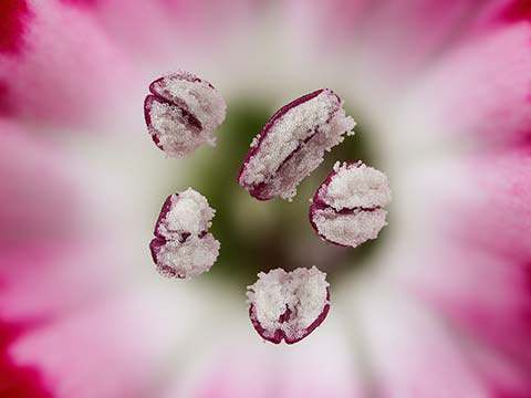
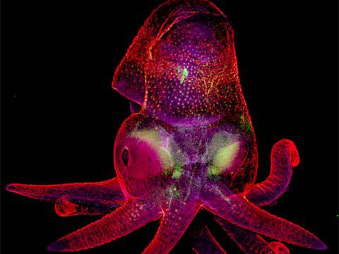
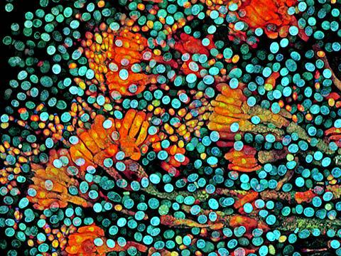
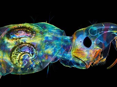
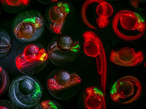
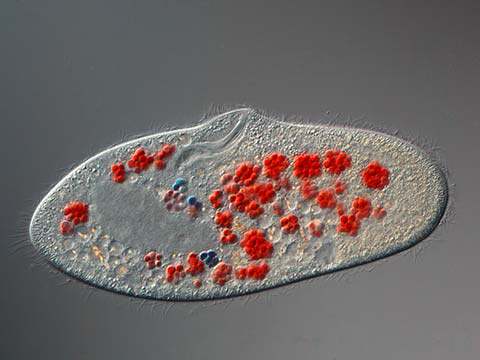
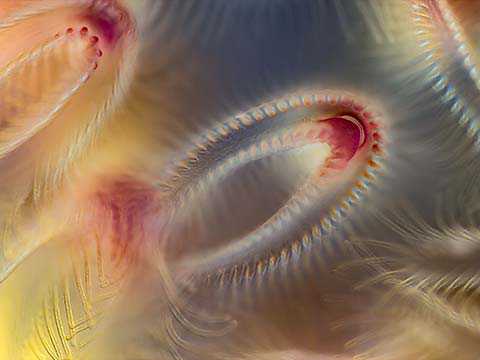
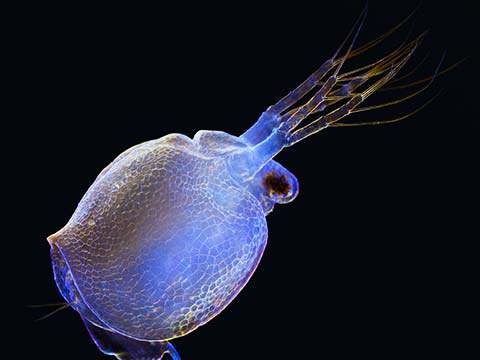
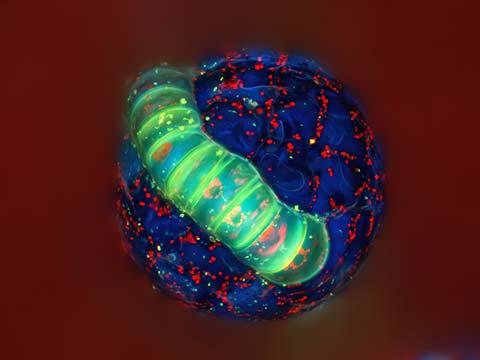
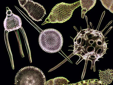
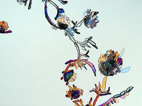
19th Place
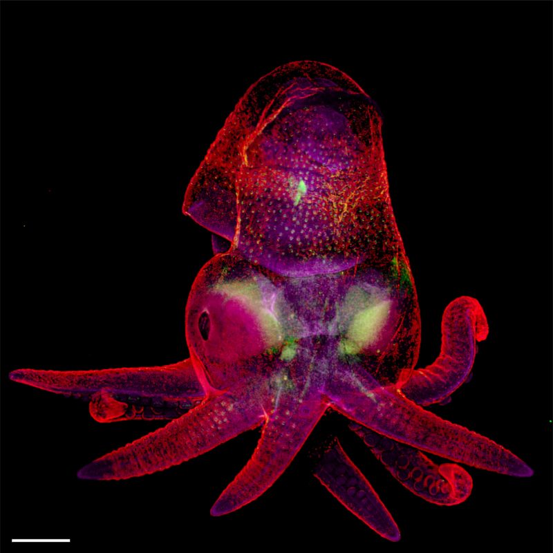
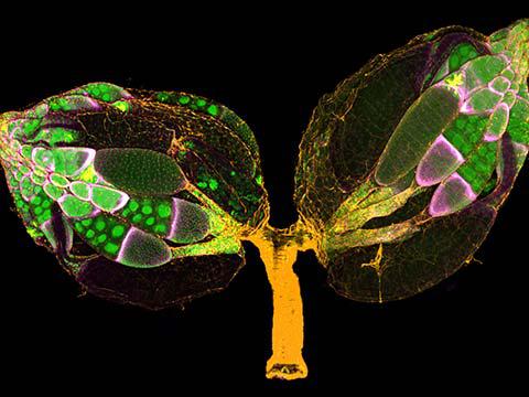
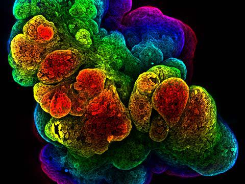
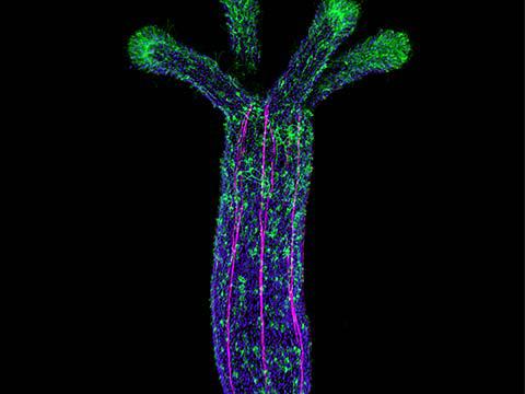
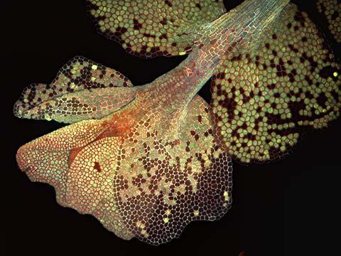
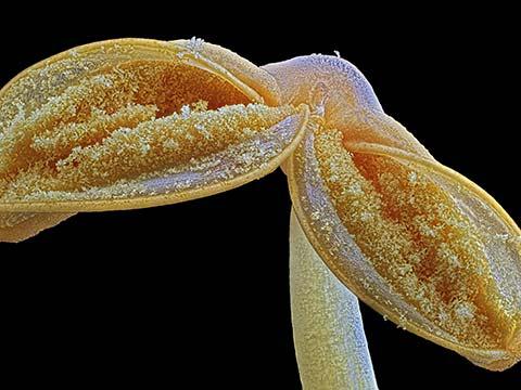
Octopus bimaculoides embryo
Martyna Lukoseviciute Dr. Carrie Albertin
- Affiliation
- University of Oxford
Weatherall Institute of Molecular Medicine
Oxford, Oxfordshire, United Kingdom
- Technique
- Confocal, Image Stitching
- Magnification
- 5x (Objective Lens Magnification)
In Their Own Words
A Q&A with Nikon Small World winner Martyna Lukoseviciute.
What is the subject matter of your winning image and why did you choose this image?
It’s an octopus embryo. Octopus is my favorite sea animal. I am fascinated by its intelligence, camouflage abilities and social interactions, but it is very different from mammals. Therefore, it is important to study its development, especially the nervous system development that can hold answers regarding the conserved and basic features of intelligent animals. This image is very beautiful and also educates people on the importance of understanding marine animal development.
What are the special techniques and/or challenges faced in creating this photomicrograph?
The biggest challenge was mounting the embryo onto the slide, because of its relatively big size. I needed to use a specialized slide with a little groove and build little walls around the sample to put the coverslip on top of it to avoid squashing the embryo. Also, because of its size, I needed to use a 5x objective and acquire multiple files to cover the whole area of the sample. This took a long time but was really worth it!
What is your primary line of work?
I am in my last year of my Ph.D. in developmental biology. I am researching neural crest specification and early differentiation controlled by epigenetic mechanisms in the zebrafish embryo.
How long have you been taking photographs through a microscope? What first sparked your interest in photomicrography?
I started taking photographs through a microscope in 2015 during my bachelor’s degree, when I was working on frog embryos. However, I have only started using high-resolution confocal microscopes during my DPhil at the University of Oxford for my work with zebrafish embryos. Seeing is believing! I was mesmerized by the beautiful structures hidden from our eyes and once I found a way to remove this barrier by using microscopes, I completely fell in love with it. I love sharing and engaging people outside the scientific community by showing my microscopy pictures of different species' embryos.
Do you tend to focus your microscopy toward a specific subject matter or theme? If so, why?
Usually, my microscopy is focused on embryo development. Nothing fascinates me more than unraveling the secrets of embryo development and comparing different species.
Why did you enter the Nikon Small World Photomicrography competition? What do you think of the competition?
I entered this competition because every year I am excited to see the winning images and learn something new about both the world and different techniques. I was excited to share my octopus image with people like me — excited to learn something new and admire the hidden beauty. As a scientist, it’s relevant to me to share images of such embryos, showing how important it is to study and compare the basic principles of embryo development. Hopefully, images like this can bring the attention back to basic science, marine biology and evolution.
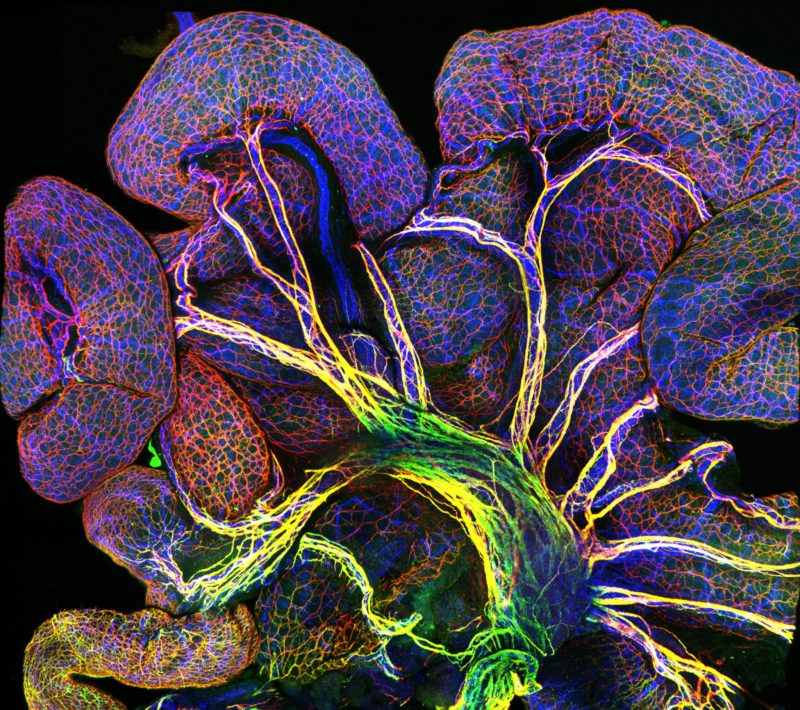
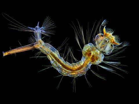
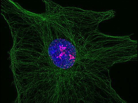
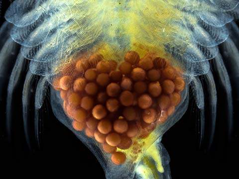
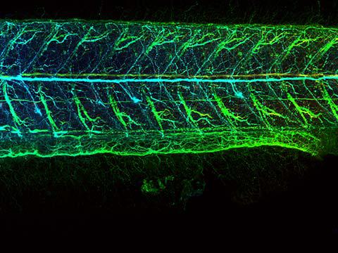
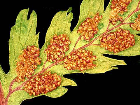
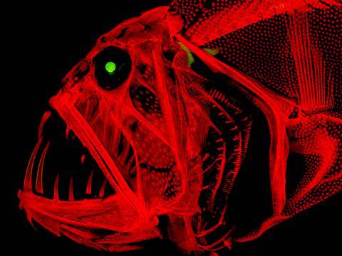
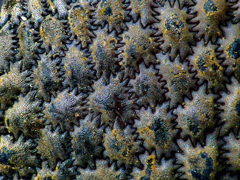
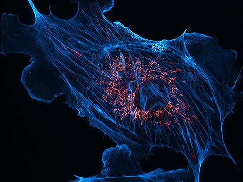
Honorable Mention
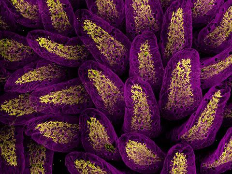
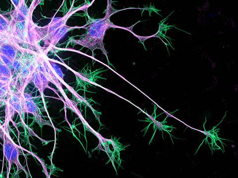
Endothelial cells in intestine of an 18.5-day old mouse embryo. Blood vessels and intestinal lining in blue; nerves in red and green.
Nathan Burns
- Affiliation
- National Institutes of Health (NIH)
National Heart, Lung, and Blood Institute
Bethesda, Maryland, USA
- Technique
- Confocal
- Magnification
- 10x (Objective Lens Magnification)
Top 20
Honorable Mentions

Images of Distinction


Judges
Dr. Denisa Wagner
Edwin Cohn Professor of Pediatrics at Harvard Medical School and the Head of the Wagner Lab Harvard Medical School and Boston Children’s Hospital
Dr. Rita Strack
Senior Editor Nature Methods
Tom Hale
Staff Writer IFLScience
Ben Guarino
Science Reporter The Washington Post