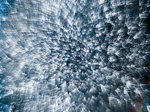
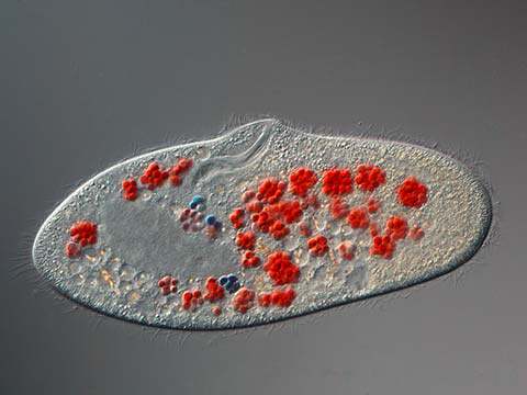
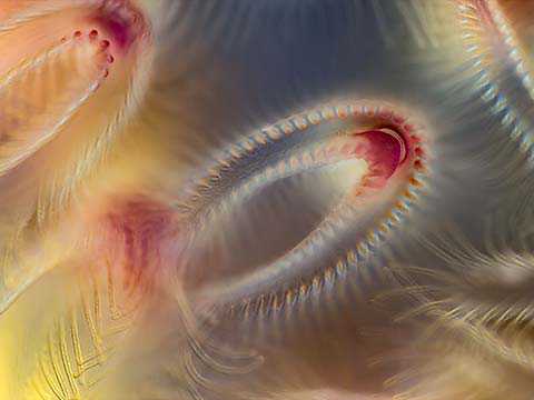
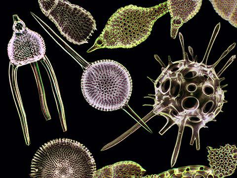
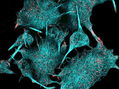
Nikon Small World veteran Dr. Igor Siwanowicz took second place in the 2019 Photomicrography Competition for his composite image of three single-cell freshwater protozoans, sometimes called "trumpet animalcules.” He used confocal microscopy to capture the detail of the cilia, tiny hairs used by the animals for feeding and locomotion.
2019 Photomicrography Competition
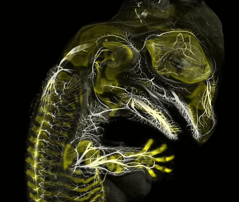
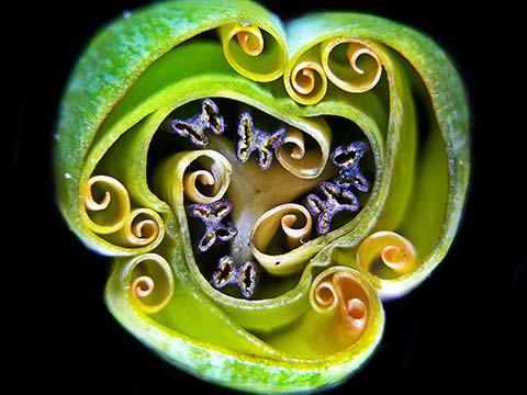
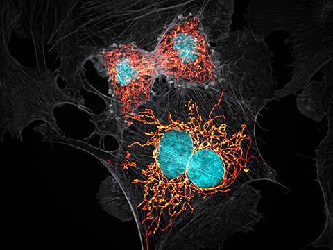
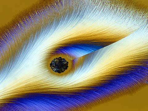
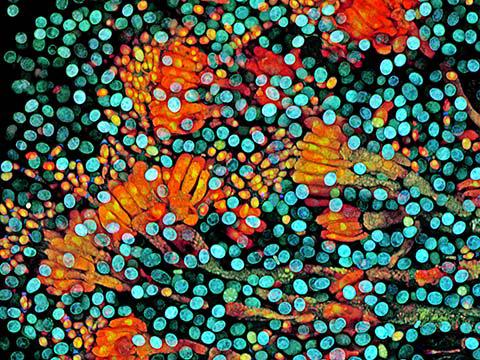
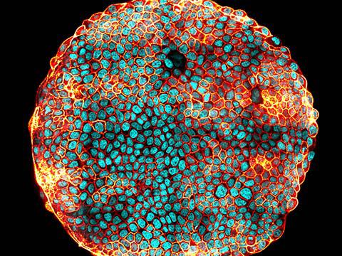
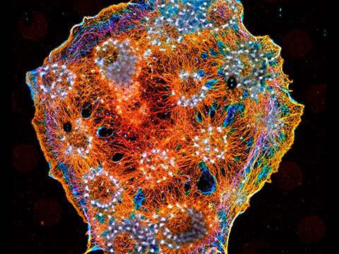
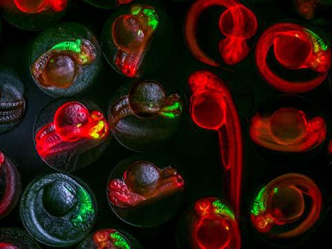
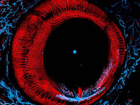
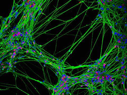
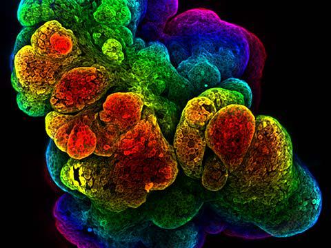
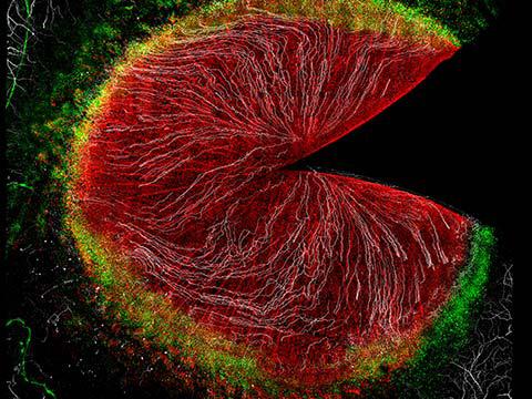
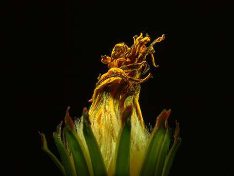
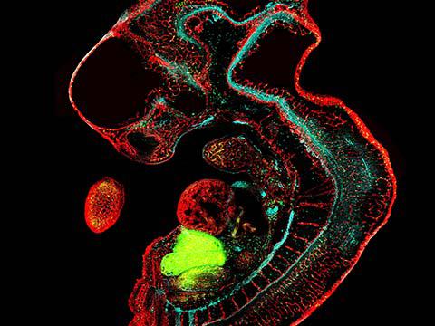
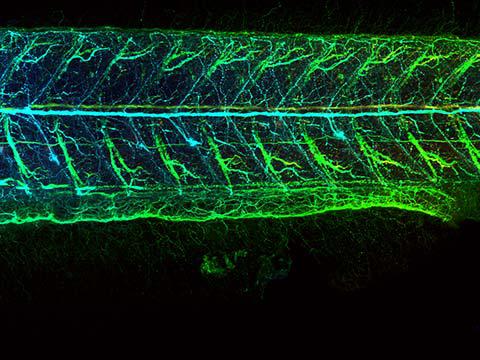
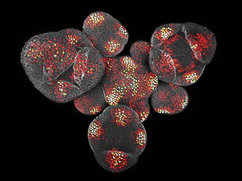
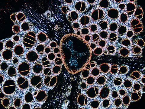
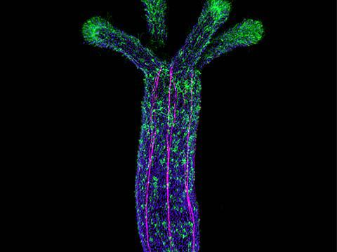
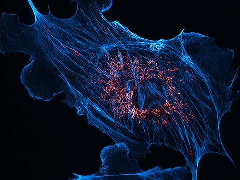
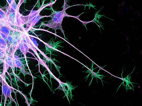
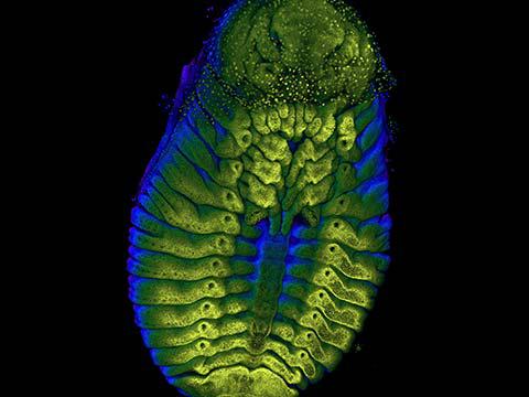
3rd Place
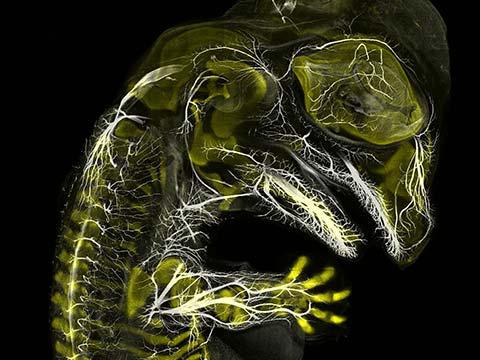
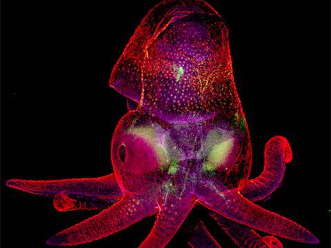
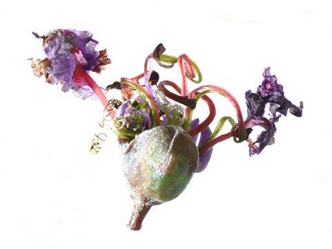
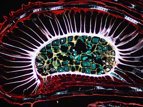
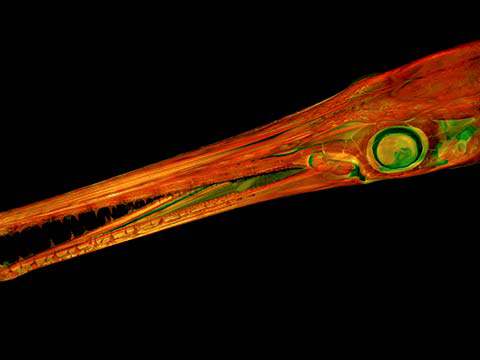
Alligator embryo developing nerves and skeleton
Daniel Smith Paredes Dr. Bhart-Anjan S. Bhullar
- Affiliation
- Yale University
Department of Geology and Geophysics
New Haven, Connecticut, USA
- Technique
- Immunofluorescence
- Magnification
- 10x (Objective Lens Magnification)
In Their Own Words
A Q&A with Nikon Small World winner Daniel Smith Paredes.
What is the subject matter of your winning image and why did you choose this image?
It’s an embryo of an American alligator, at around 20 days of development, stained to label the developing nerves (white) and skeleton (yellow). I thought it looked cool, and it showed how intricate the nerves can be even early in development, and how they relate spatially to the skeleton.
What are the special techniques and/or challenges faced in creating this photomicrograph?
The most difficult task was to be able to image something this size. It’s actually composed of many thousand individual pictures merged together.
What is your primary line of work?
Evolutionary developmental biology, comparative anatomy and embryology. Studying the way vertebrates have evolved by comparing their embryonic development and how it is similar and how is different between different groups of animals.
How long have you been taking photographs through a microscope?
Three-four years. I was inspired by the ability to look into the anatomical structures in developing embryos.
Do you tend to focus your microscopy toward a specific subject matter or theme? If so, why?
My interest is directed mostly towards embryonic anatomy and development.
Why did you enter the Nikon Small World Photomicrography competition? What do you think of the competition?
I was encouraged by a friend who told me I should give it a try, since my samples look good and also because they represent some groups of animals mostly understudied or photographed in micro scale.
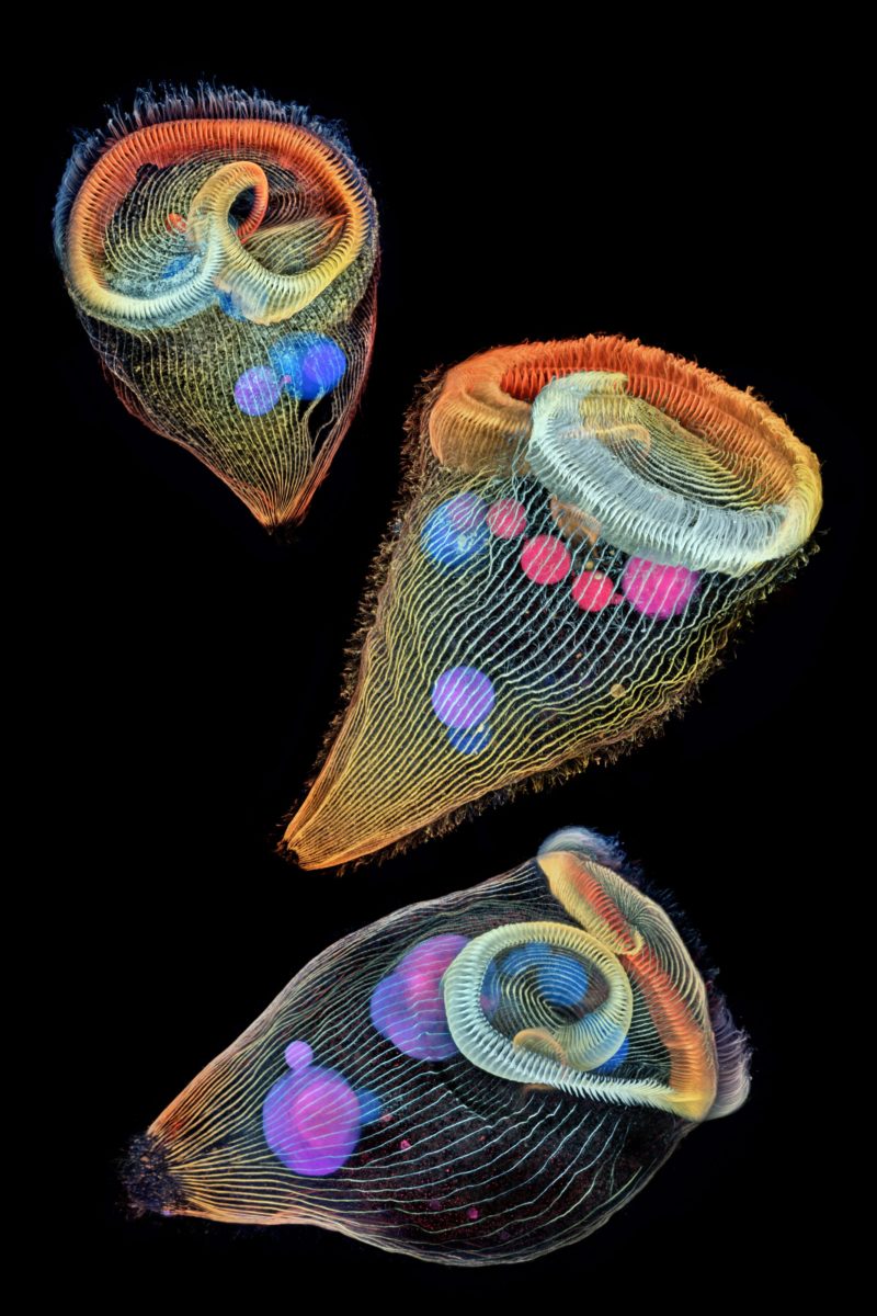
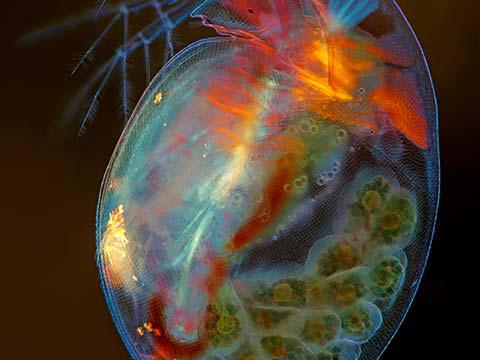
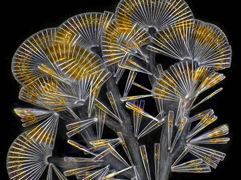
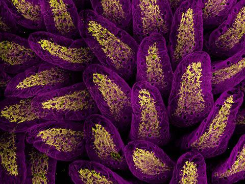
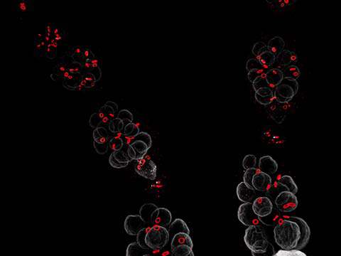
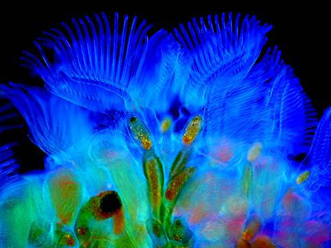
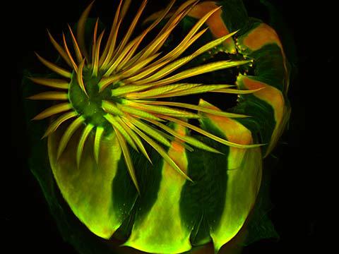
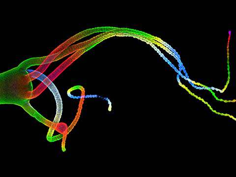
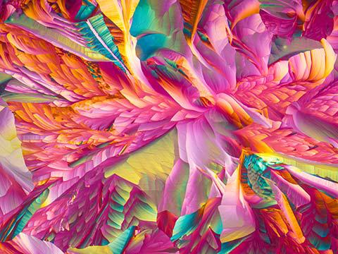
2nd Place
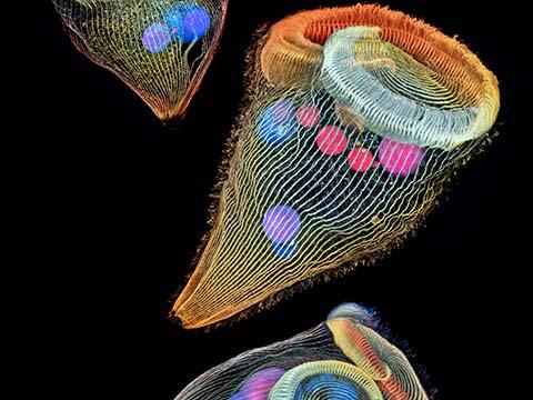
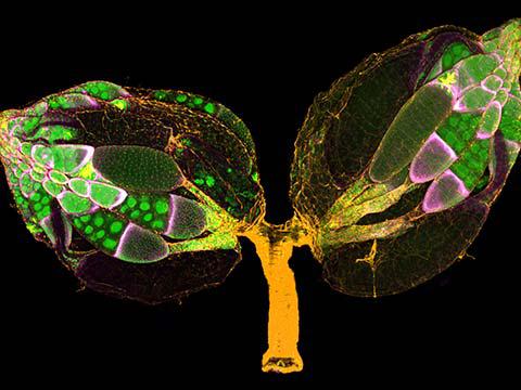
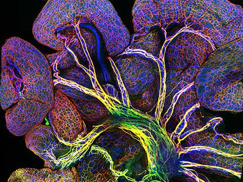
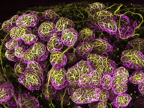
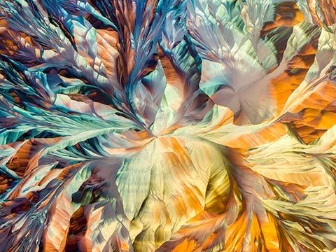
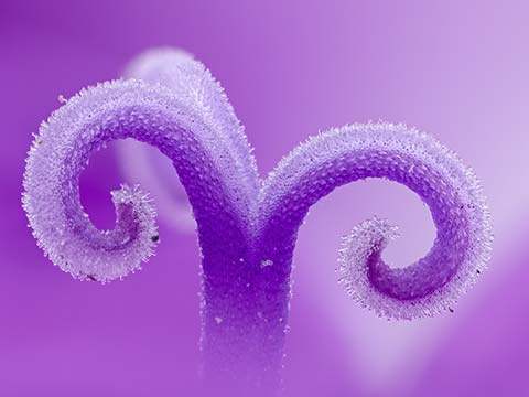
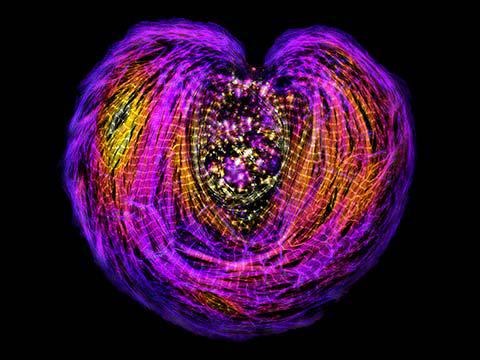
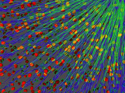
Depth-color coded projections of three stentors (single-cell freshwater protozoans)
Dr. Igor Robert Siwanowicz
- Affiliation
- Howard Hughes Medical Institute (HHMI)
Janelia Research Campus
Ashburn, Virginia, USA
- Magnification
- 40x (Objective Lens Magnification)
In Their Own Words
A Q&A with Nikon Small World winner Dr. Igor Siwanowicz.
What is the subject matter of your winning image and why did you choose this image?
The image, a composite of three depth color-coded projections, shows single-cell freshwater protozoans, stentors (sometimes called “trumpet animalcules”). Stentors were for quite a while on my list of potential imaging subjects, but, although present in local ponds, I could never collect a sufficient amount to try various fixation and staining protocols. Last spring, we had two Ph.D. students visiting our advanced imaging center with a goal of visualizing mitochondrial dynamics in those protozoans. I simply asked them to kindly let me use their leftover stocks after they were done with them. Stentors were featured in the Nikon Smal World contest multiple times, but I don’t recall seeing any images of the protists obtained with a confocal imaging technique. I thought that made the image somewhat unique.
What are the special techniques and/or challenges faced in creating this photomicrograph?
I used antibodies against acetylated tubulin to visualize the cilia that those tiny animals (0.5 mm long) use for feeding and locomotion, and DAPI, a DNA-binding compound, to image the nuclei that form a long strand reminiscent of a string of pearls. Fixation of the protozans is notoriously difficult. When exposed to fixative the organisms tend to collapse into a ball of protoplasm rather than retain their natural extended form. I finally found a working protocol in a 1973 publication.
What is your primary line of work?
For the past eight years, I have been studying neuroanatomy of a dragonfly, specifically the neural circuits involved in pray interception and capture. More recently, I’m collaborating on a number of projects (in- and outside of Janelia), that involve imaging of various chunks of invertebrate anatomy.
How long have you been taking photographs through a microscope? What first sparked your interest in photomicrography?
About 10 years. I was fascinated with nature since before I remember; my parents are biologists and I grew up surrounded by textbooks. I enjoyed browsing through the illustrations and photographs long before I learned how to read. It wasn’t until 16 years ago, at the age of 26, that I bought my first camera and found myself on the supply side of nature photography, with special focus on macro technique. Photomicrography is a logical continuation of photography and allows me an even more intimate perspective of my “models”.
Do you tend to focus your microscopy toward a specific subject matter or theme?
I’m enamored with invertebrate morphology; usual evolutionary restraints don’t seem to apply within the realm of tiny animals, which is evident in the abundance and variety of often grotesque and utterly alien forms. Microscopy allows me to see beyond the cuticle, explore the baroque arrangement of muscle fibers or intricate fractal-like network of neurons.
Why did you enter the Nikon Small World Photomicrography competition? What do you think of the competition?
Many scientists share an appreciation of beauty and are fully aware of the aesthetic aspects of their research. The Nikon Small World Contest was conceived with such people in mind. Images are rewarded for the artistic merit and visual aspects on par with and often above their scientific importance; that definitely grants the contest a broad appeal among non-experts and contributes to redeeming the image of science as a somber, wonder-less, unexciting affair utterly unintelligible for a layperson.
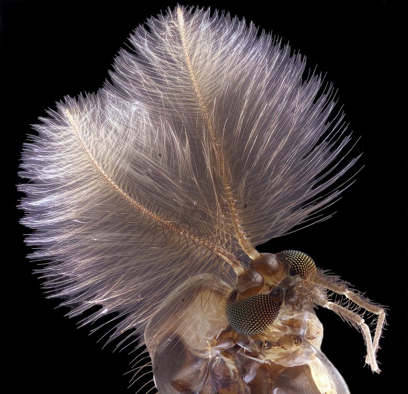
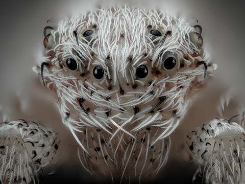
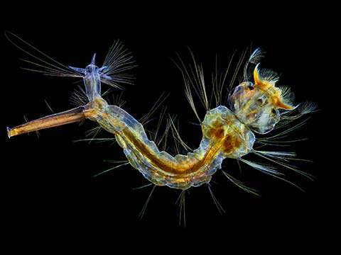
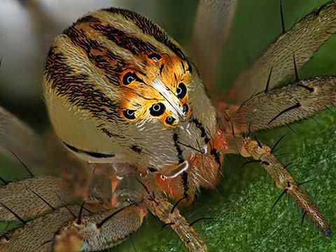
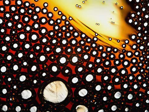
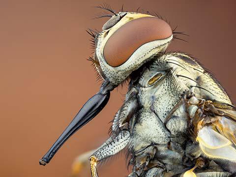
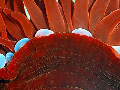
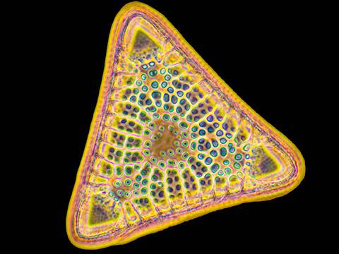
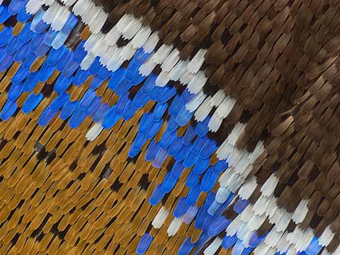
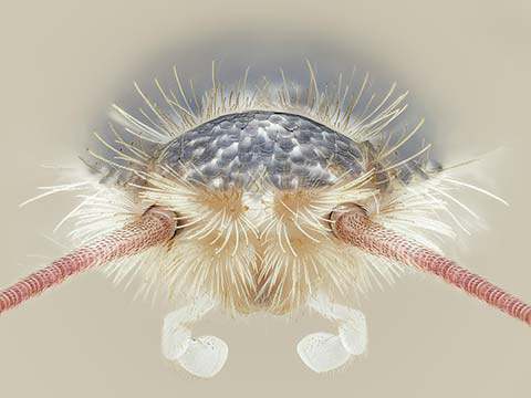
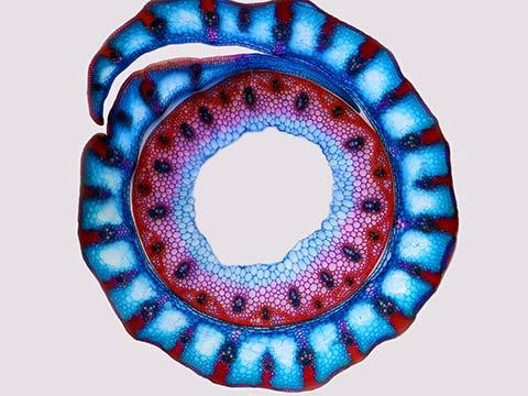
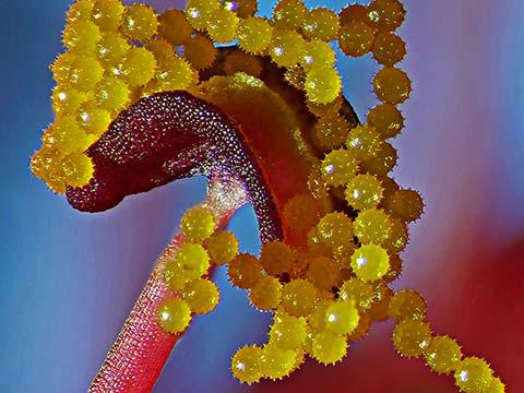
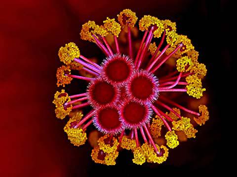
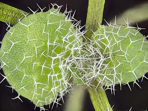
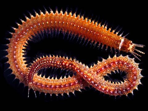
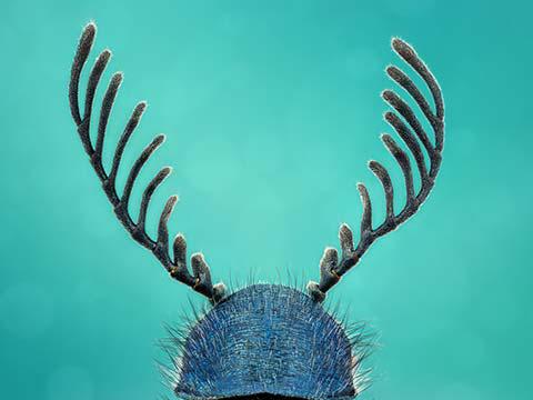
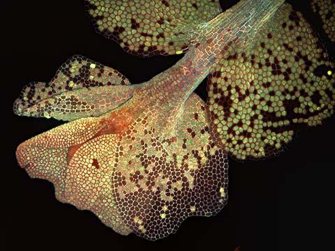
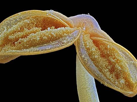
4th Place
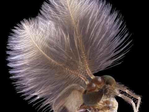
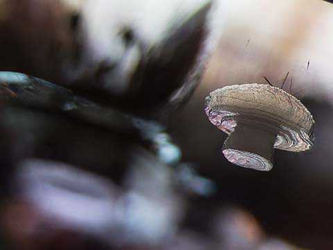
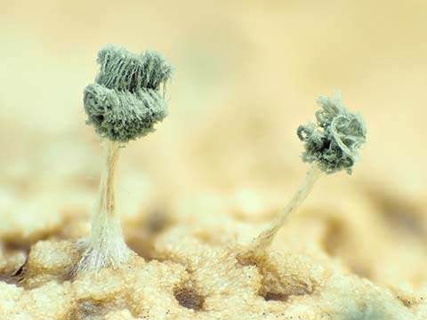
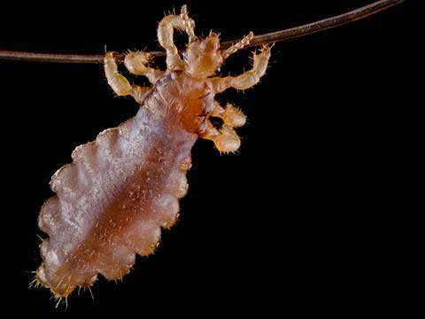
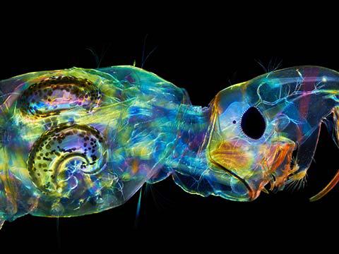
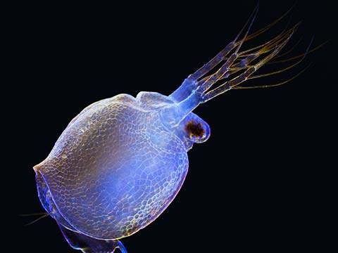
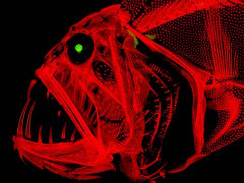
Male mosquito
Jan Rosenboom
- Affiliation
- University of Rostock
Rostock, Mecklenburg Vorpommern, Germany
- Technique
- Focus Stacking
- Magnification
- 6.3x (Objective Lens Magnification)
In Their Own Words
A Q&A with Nikon Small World winner Jan Rosenboom.
What is the subject matter of your winning image and why did you choose this image?
My image shows a male mosquito. I chose to submit it because I really liked its composition. The huge antennae make a great contrast to the mosquito's green eyes. Only male mosquitos have such big antennae as a form of attraction to the other sex. I also liked the contrast between the beauty of the mosquito and how we just kill them without caring how they look like.
What are the special techniques and/or challenges faced in creating this photomicrograph?
A particular challenge was the movement of the huge antennae the mosquito has. If there is any movement, the different layers in the focus stacking process will not align. Therefore, I even had to pay attention to my breathing to not get the antennae moving.
How long have you been taking photographs through a microscope? What first sparked your interest in photomicrography?
Five years, since I was 16. I got into photomicrography accidentally when I got an old Zeiss microscope (1960) and started experimenting with what you could do with it using modern technology. Over the years, I perfected my methods and improvised a lot of special low-budget equipment in order to be able to compete with microscopes costing thousands and thousands of dollars. I am now 22 and it still fascinates me how it is possible to produce beautiful images with quite basic equipment when you are experienced enough.
Do you tend to focus your microscopy on a specific subject matter or theme? If so, why?
I tend to focus on insects. It amazes me how seemingly mundane species such as a common housefly or a mosquito look like beautiful creatures from space when you just look at them with enough detail.
Why did you enter the Nikon Small World Photomicrography competition?
I decided to enter the competition because I wanted to show how you can compete with scientific institutions using very expensive microscopes with DIY techniques and enough experience and creativity.
Top 20
Honorable Mentions

Images of Distinction

Judges
Dr. Denisa Wagner
Edwin Cohn Professor of Pediatrics at Harvard Medical School and the Head of the Wagner Lab Harvard Medical School and Boston Children’s Hospital

Dr. Wagner has dedicated her career to the fields of vascular cell biology and the causes of inflammation and blood clots. For many years, her laboratory’s research has focused on adhesion molecules (cell adhesion molecules help cells stick to each other and to their surroundings) and their function in normal physiology and in pathological situations. One of her labs’ current interests is the biology of neutrophil extracellular traps (networks of extracellular nuclear DNA) and the study of their production using time-lapse microscopy.
Dr. Rita Strack
Senior Editor Nature Methods
Dr. Strack has been an editor at Nature Methods since November of 2014. Her primary areas of coverage for the journal are imaging, microscopy, and probes, but her interests and expertise also extends to molecular biology, structural biology, and biophysics. She attended the University of Chicago, where she earned her Ph.D. in Biochemistry in 2010. Strack did a postdoctoral fellowship at Weill Cornell College of Medicine, where she spent countless hours on the microscope getting beautiful images to better understand the toxic RNAs associated with Fragile-X tremor, an adult-onset neurodegenerative disease.
Tom Hale
Staff Writer IFLScience
Hale is a London-based journalist at beloved popular science publication IFLScience. He is an experienced science writer, researching and sharing insights on everything from new species and biomedical breakthroughs to climate change and viruses. He has also written on art in culture, his work appearing in publications such as VICE’s Motherboard and FACT Magazine.
Ben Guarino
Science Reporter The Washington Post
A top science writer for The Washington Post, Guarino focuses on the practice and culture of science. Before making the switch to journalism, Guarino studied bioengineering and worked at the Spine Pain Research Lab at the University of Pennsylvania. He has also worked as a freelance science journalist, an associate editor at the Dodo and a medical reporter at the McMahon Group. His work has also appeared in publications like The Verge and The Huffington Post.