2016 Photomicrography Competition
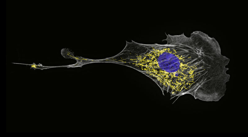
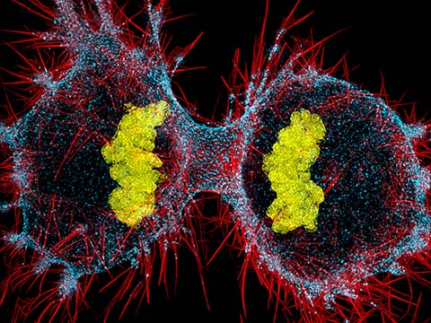
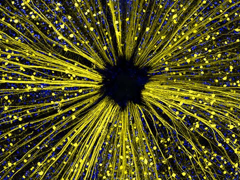
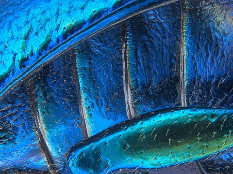
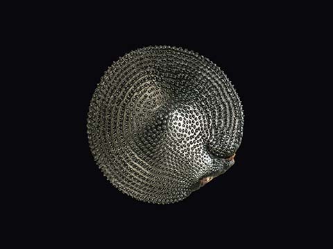
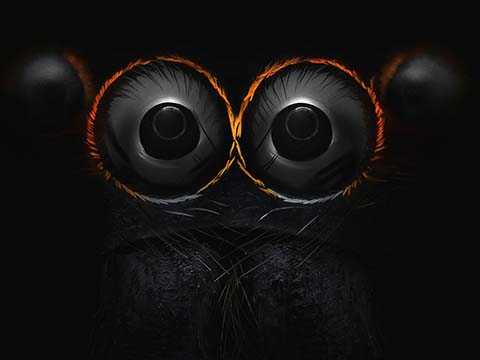
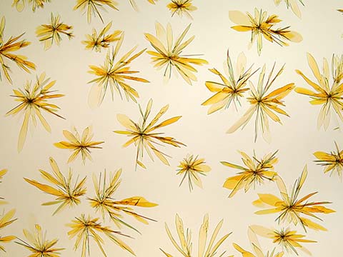
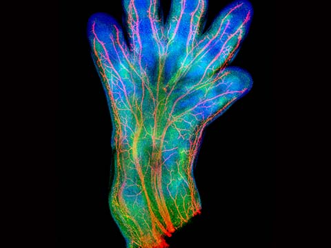
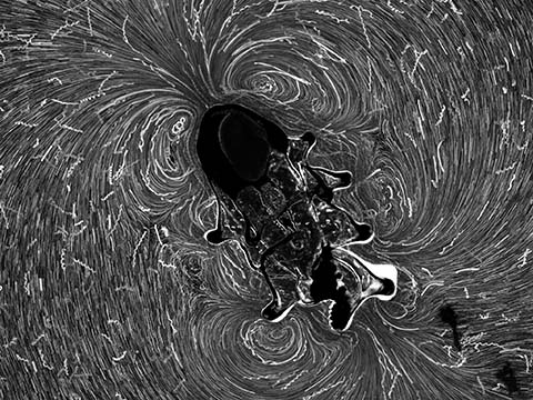
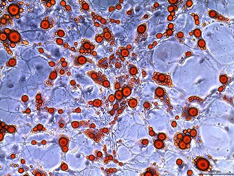
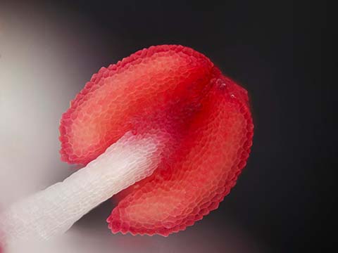
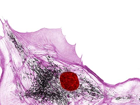
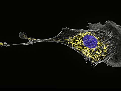
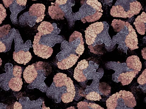
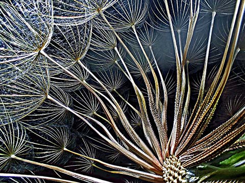
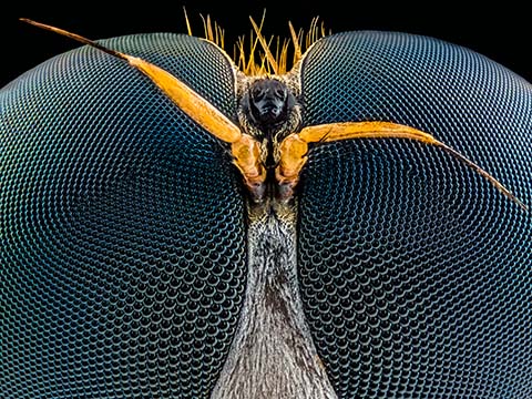
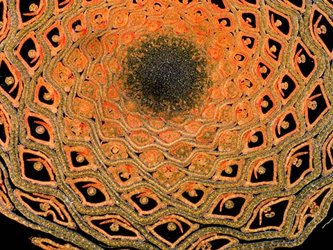
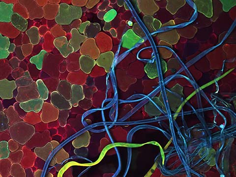
Image of Distinction
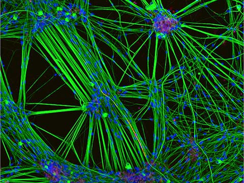
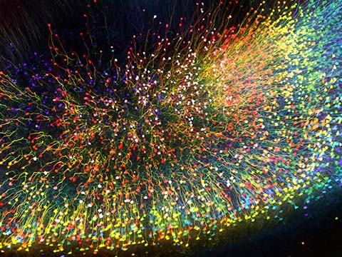
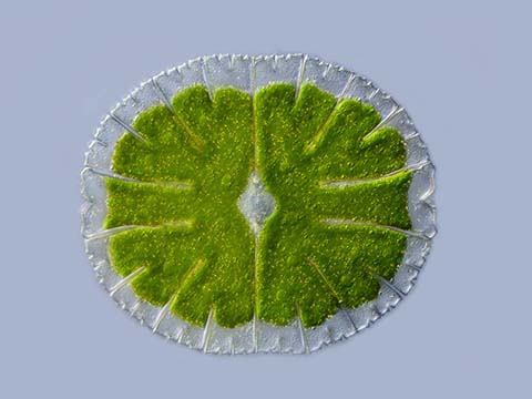
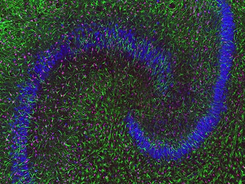
Actin (white), mitochondria (yellow), and DNA (blue) in a bovine pulmonary artery endothelial cell
Dr. Talley J. Lambert
- Affiliation
- Harvard Medical School
Department of Cell Biology
Boston, Massachusetts, USA
- Technique
- Structured Illumination Microscopy
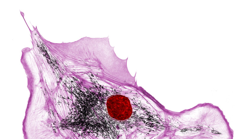
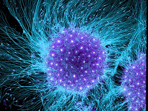
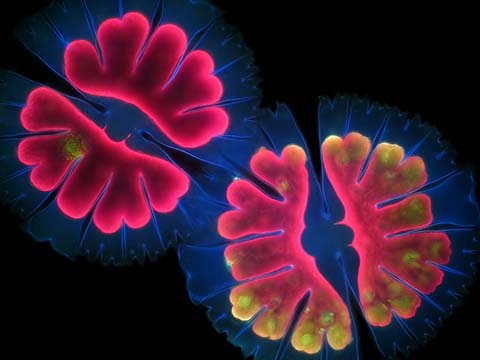
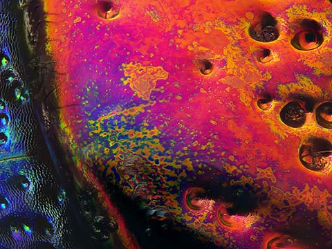
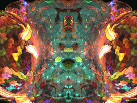
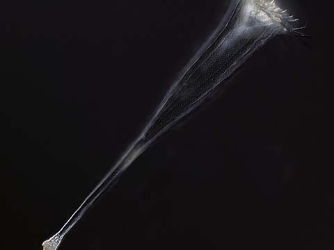
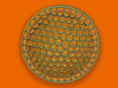
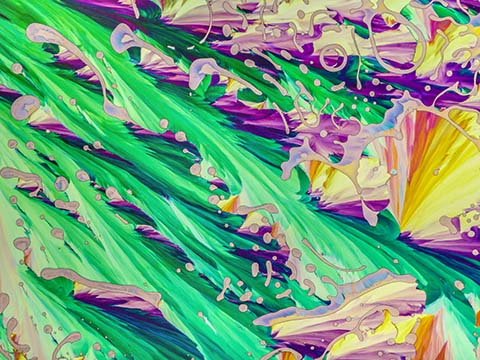
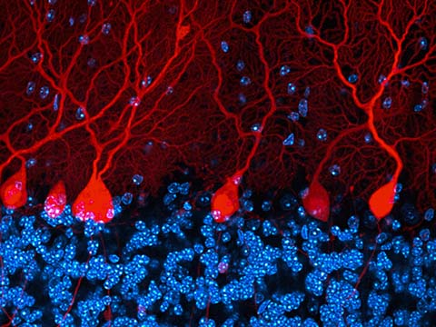
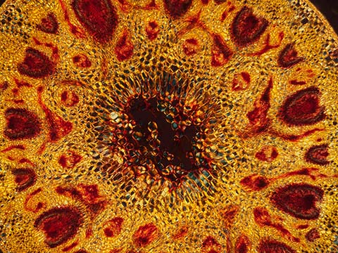
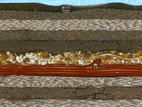
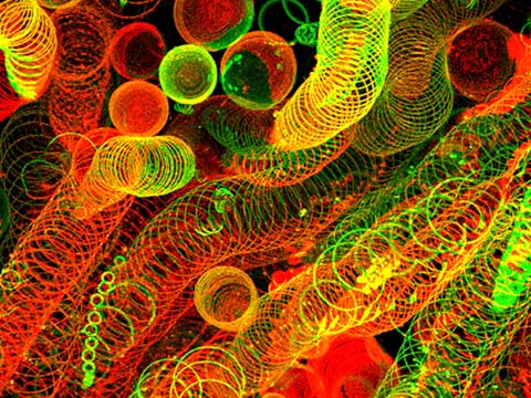
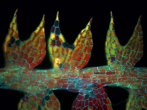
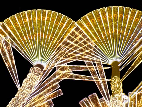
Image of Distinction
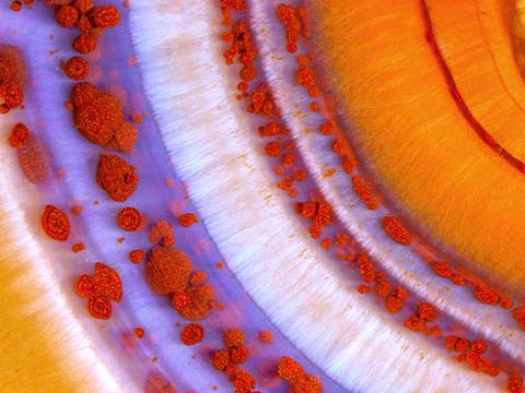
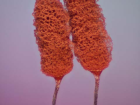
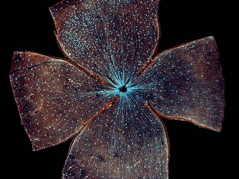
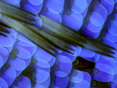
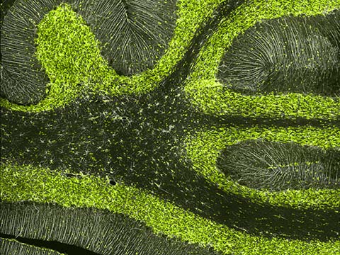
Actin (pink), mitochondria (black), and DNA (red) in a bovine pulmonary artery endothelial cell
Dr. Talley J. Lambert
- Affiliation
- Harvard Medical School
Department of Cell Biology
Boston, Massachusetts, USA
- Technique
- 3D Structured Illumination Microscopy
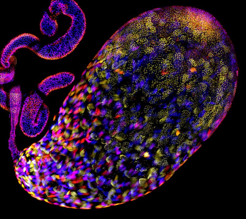
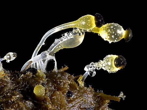
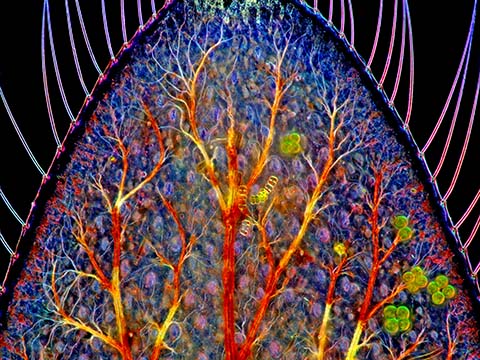
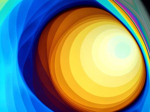
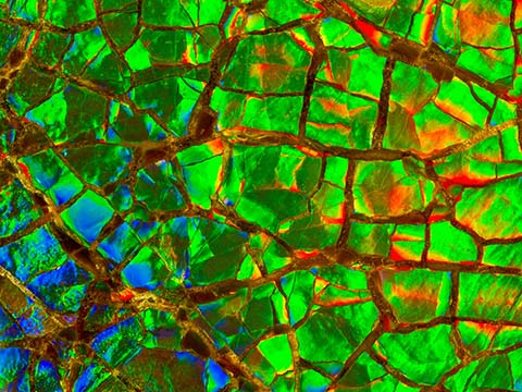
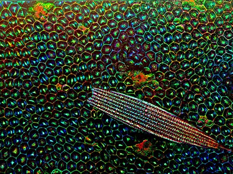
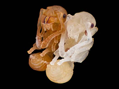
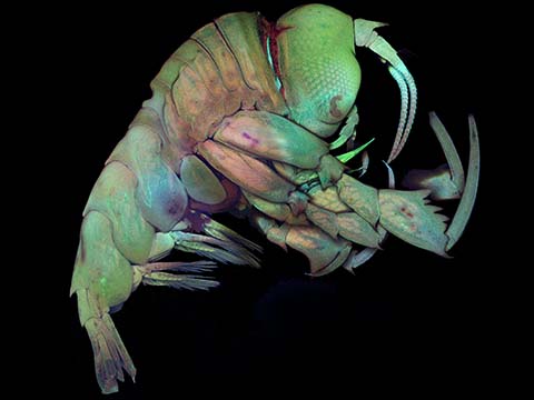
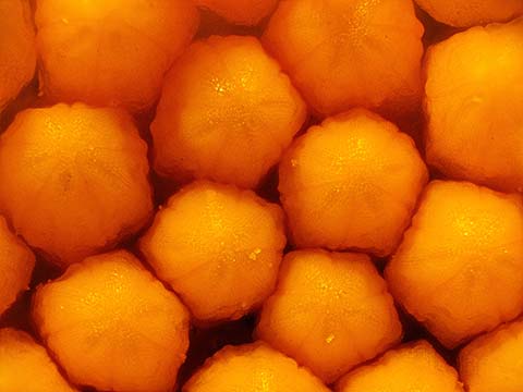
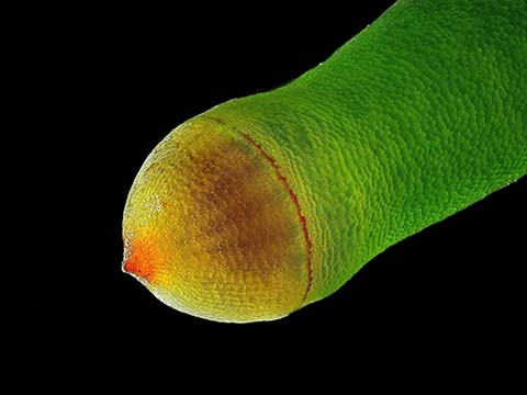
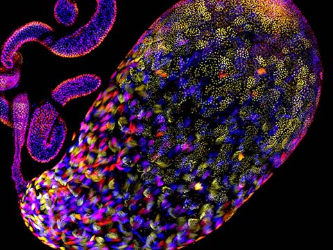
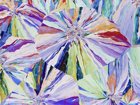
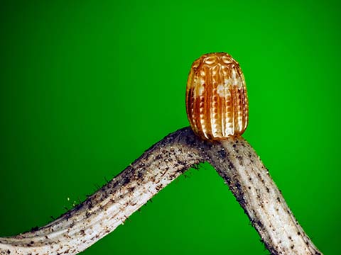
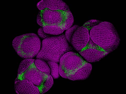
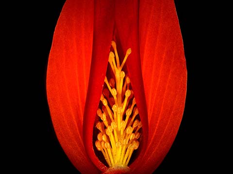
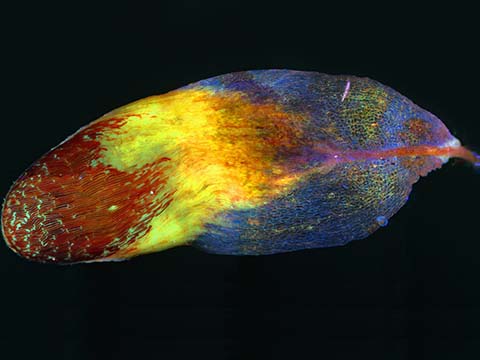
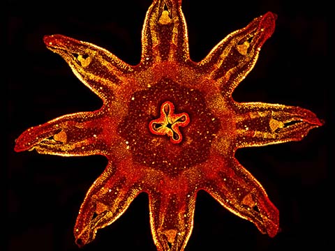
Image of Distinction
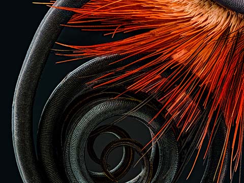
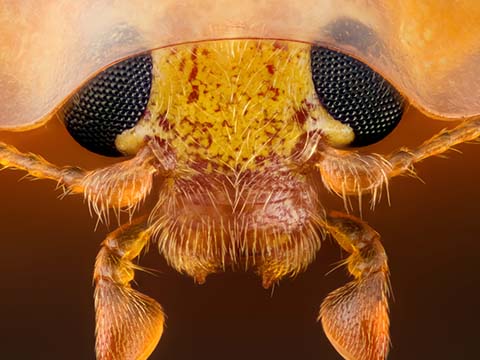
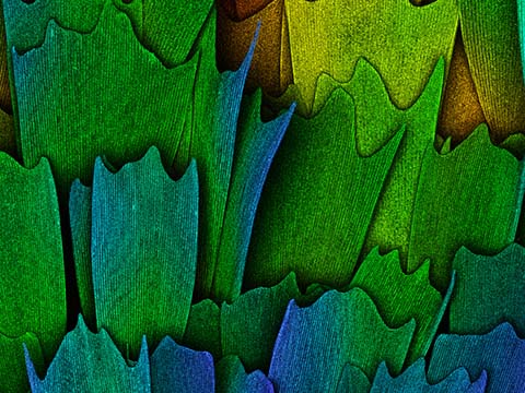
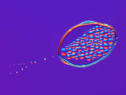
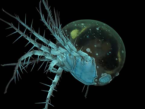
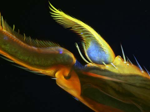
Testis of a fruit fly (Drosophila pseudoobscura) stained for DNA and pseudocolored for depth, showing the stages of sperm development
Christopher Large
- Affiliation
- University of Utah
Department of Biology
Salt Lake City, Utah, USA
- Technique
- Confocal
- Magnification
- 60x
Top 20
Honorable Mentions

Images of Distinction
Judges
Dr. Clare Waterman
Distinguished Investigator at the Laboratory of Cell and Tissue Morphodynamics National Institute of Health (NIH)
Clare Waterman graduated from the Mount Holyoke college with a B.A. in biochemistry in 1989, received an M.S. in exercise science in 1991 from the University of Massachusetts, and received her Ph.D. in cell biology from the University of Pennsylvania in 1995. Prior to joining the NHLBI, she spent 9 years as a professor in the Department of Cell Biology at the Scripps Research Institute in La Jolla, CA. Dr. Waterman is a NIH Distinguished Investigator and has received numerous awards and honors for her work, including the Arthur S. Flemming Award for Public Service (Basic Science) from George Washington University.
Dr. Waterman has made fundamental advances in the understanding of cytoskeletal interactions and has authored or coauthored more than 90 papers. She currently serves on the editorial boards of Current Biology and Journal of Microscopy. Dr. Waterman is a member of the American Society for Cell Biology, Royal Microscopical Society, Biophysical Society, and is a Council Member of Gordon Research Conferences Organization.
Dr. Brian Mitchell
Associate Professor in Cell and Molecular Biology Northwestern University Feinberg School of Medicine
Brian Mitchell is an Associate Professor in Cell and Molecular Biology at Northwestern University Feinberg School of Medicine in Chicago IL. He is also a member of the Lurie Cancer Center at Northwestern.
Brian graduated from the University of San Diego with a B.A. in biology and marine studies in 1993, and received his Ph.D. in neurobiology from the University of North Carolina, Chapel Hill, in 2001. He is currently a member of the American Society of Cell Biologists, Society of Developmental Biologists and Genetics Society of America. In 2016 he was the Nikon Fellow at the Marine Biological Labs in Woods Hole MA. His work focuses on using microscopic approaches to address a broad range of questions in cell and developmental biology.
Dr. Joe Hanson
Creator and host of PBS Digital Studios’ science education show “It’s Okay To Be Smart”

Dr. Joe Hanson (@jtotheizzoe) is a biologist, science writer, and the creator and host of PBS Digital Studios’ science education show It’s Okay To Be Smart, which takes a fun-loving look at the world of science through the lens of pop culture, art, and comedy.
Joe’s award-winning writing has been published in WIRED, Nautilus, ScientificAmerican.com, and “It’s Okay To Be Smart” has been nominated for IAWTV and Streamy Awards for excellence in online video.
Rachel Link
Producer National Geographic
Rachel Link shares the work of talented filmmakers from around the world curating content for National Geographic’s Short Showcase. She is excited about the ways in which video can help communicate scientific research and advancement. Her background is in educational video production and has a degree in Media Studies from Scripps College.
Eric Clark
Florida State University’s National High Magnetic Field Laboratory (NHMFL)
Eric Clark has been with Florida State University’s National High Magnetic Field Laboratory (NHMFL) since 1999. Eric serves as webmaster and applications coordinator for the Office of Research and Optical Microscopy division at the NHMFL. Prior to working at FSU, Eric was a surgical nurse and wound care specialist for ten years.