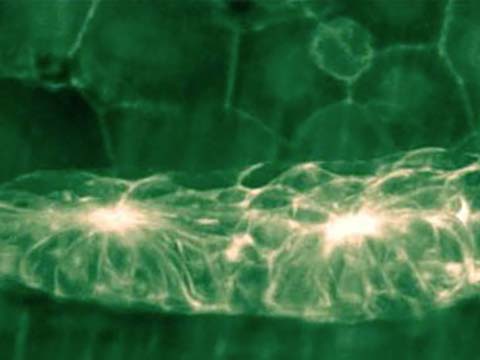
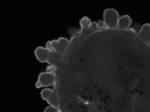
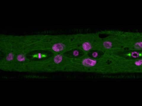
Collecting duct of three different embryonic mouse kidneys growing in ex vivo culture systems and captured every 20 minutes for 2 days. The kidneys express a nuclear-localized Green Fluorescent Protein which has been pseudocolored in three different colors and overlaid. The purpose of the overlay is to highlight the highly conserved branching behavior of these three different kidneys. The kidneys were cultured separated and digitally combined.
2014 Small World In Motion Competition
Honorable Mention
Developing mouse embryonic kidneys
Dr. Nils Lindström
- Affiliation
- The Roslin Institute
Edinburgh, Scotland, United Kingdom
- Technique
- Fluorescence
- Magnification
- 4x
Honorable Mention
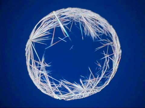
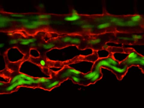
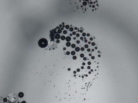
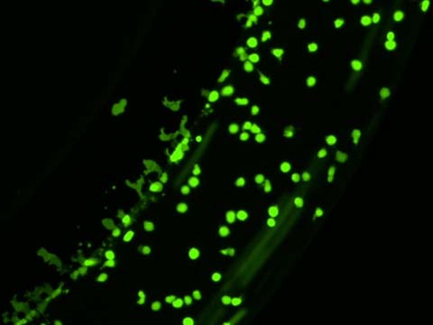
Time-lapse of a 2 day old zebrafish embryo with green erythrocytes and red blood vessels
Dr. Elliott Hagedorn
- Affiliation
- Boston Children’s Hospital
Boston, Massachusetts, USA
- Technique
- Confocal
Time-lapse of the tail region of a two day old double transgenic zebrafish embryo showing green erythrocytes and red blood vessels. Embryo was anesthetized with tricaine and mounted in low melting point agarose.
Honorable Mention
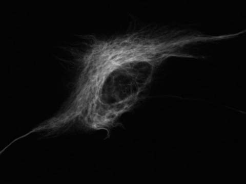
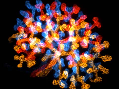
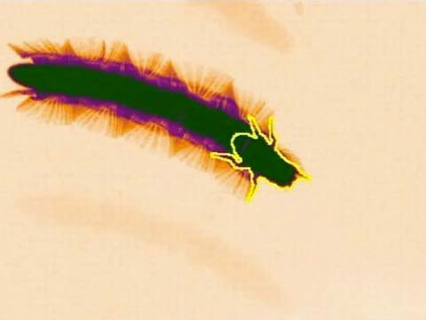
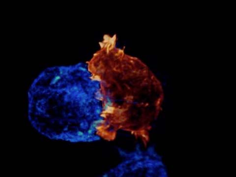
Human melanoma (cancer) cells blebbing
Dr. Jeremy Logue
- Affiliation
- National Institutes of Health (NIH)
Bethesda, Maryland, USA
- Technique
- Confocal
- Magnification
- 100x
Blebbing of human melanoma A375 cells expressing EGFP tagged F-tractin under agarose.
Top 20
Honorable Mentions
Judges
Paul Maddox
Assistant Professor and William Burwell Harrison Fellow University of North Carolina at Chapel Hill, Department of Biology
Paul S. Maddox, Ph.D. is an Assistant Professor and William Burwell Harrison Fellow, Department of Biology, University of North Carolina at Chapel Hill, North Carolina, USA. Dr. Maddox completed his Ph.D. under the mentorship of renowned cell biology microscopist E. D. Salmon at the University of North Carolina at Chapel Hill. His current research interests include using light microscopy to understand chromosome and microtubule dynamics. Dr. Maddox’s experimental philosophy mandates using the light microscope to “see” results in the most quantitative manner possible. Dr. Maddox has over 19 years of experience with light microscopy and has been an instructor in light microscopy courses around the world including Europe, South America, and at the Marine Biological Labs in Woods Hole, USA. He has published over 60 peer-reviewed papers in his career.