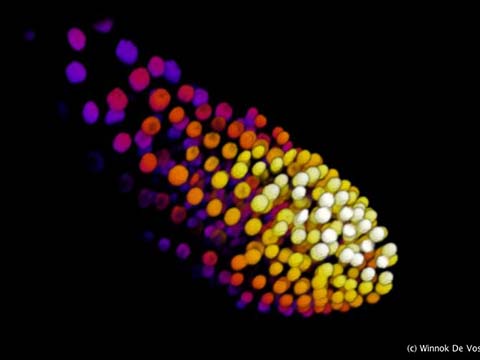
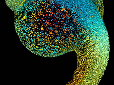
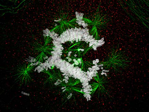
The image shows an entire zebrafish embryo at single-cell resolution and enables an insightful view of its early morphogenetic development. The embryo’s cell nuclei were fluorescently labeled and imaged with a SiMView custom light-sheet microscope. Color encodes depth in the image.
2014 Photomicrography Competition
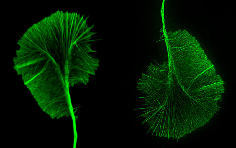
Image of Distinction
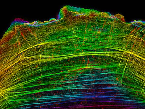
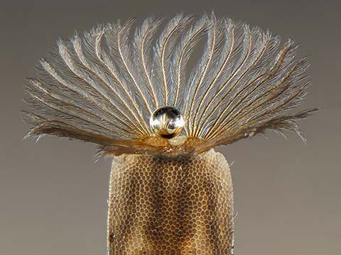
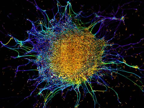
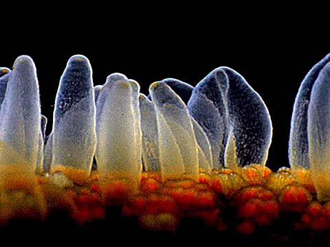
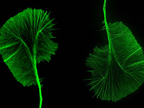
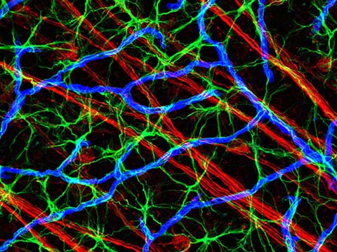
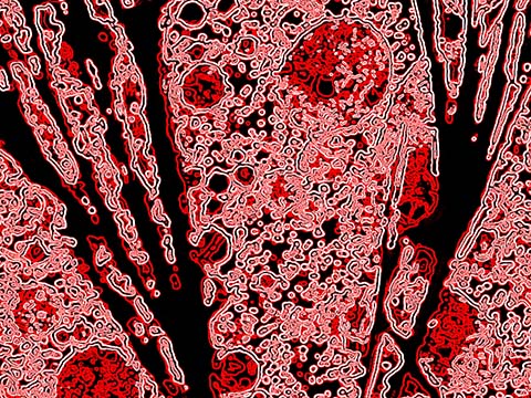
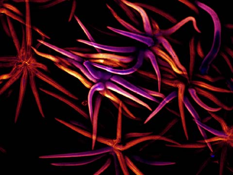
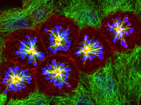
Actin meshwork of an identical growth cone
Dr. Kaoru Katoh
- Affiliation
- National Institute of Advanced Industrial Science and Technology (AIST)
Biomedical Research Inst.
Tsukuba, Japan
- Technique
- Confocal and Super-Resolution Microscopy
- Magnification
- 3000x
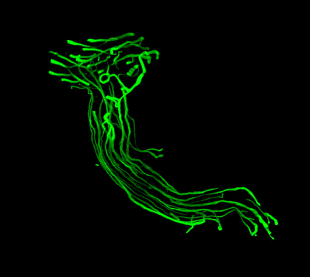
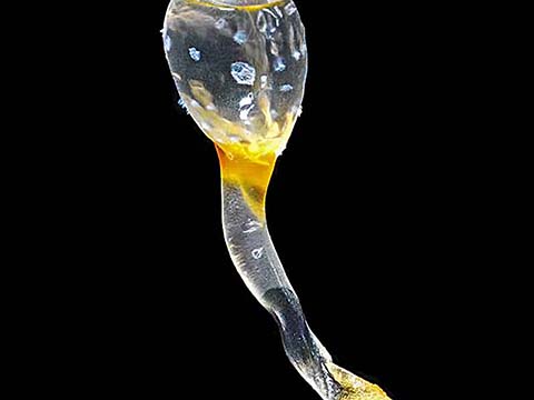
Image of Distinction
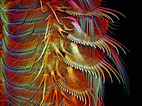
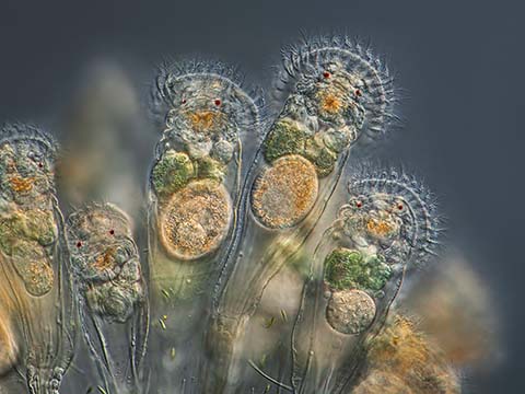
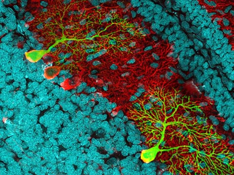
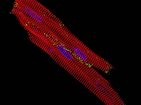
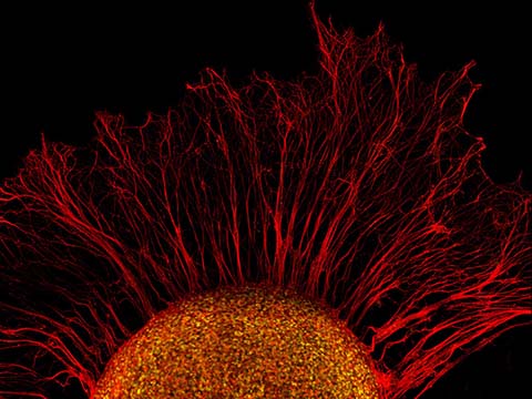
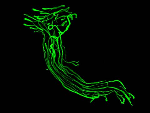
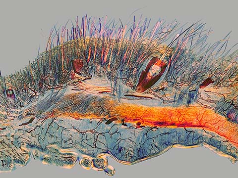
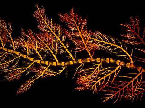
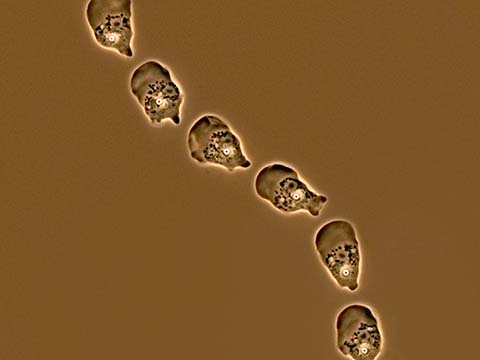
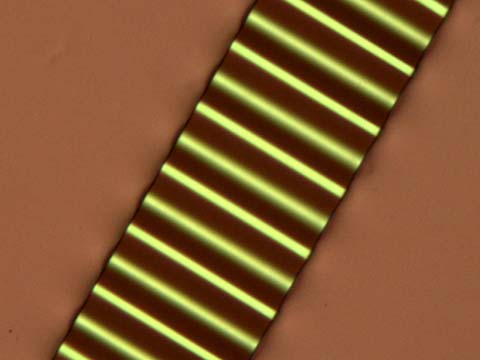
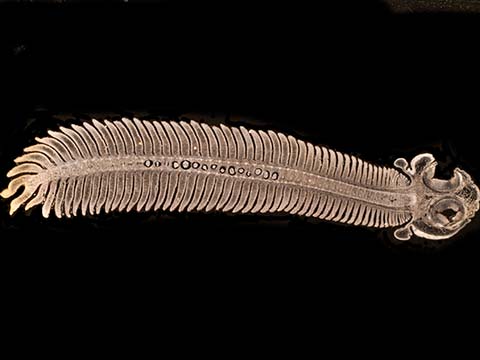
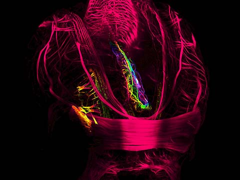
“Bioluminescence” Nerve segment in dog hind limb muscle
Cheryl Jensen
- Affiliation
- University of Missouri
Department of Ophthalmology - Neurodegenerative Diseases Research Laboratory
Columbia, Missouri, USA
- Technique
- Confocal
- Magnification
- 20x
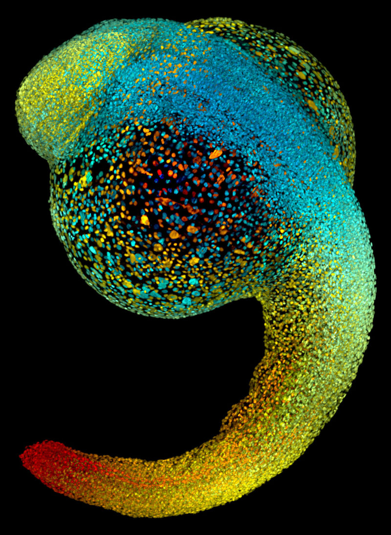
Image of Distinction
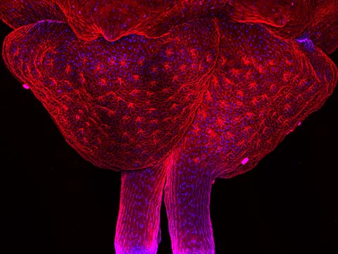
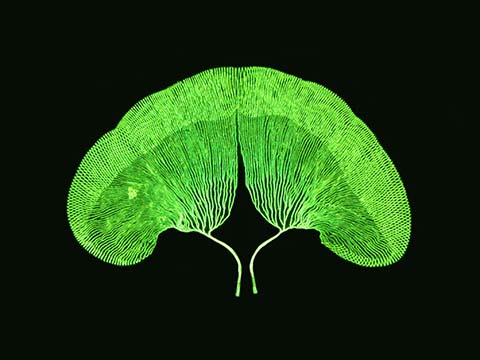
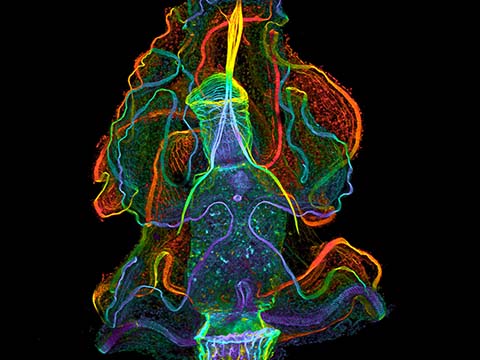
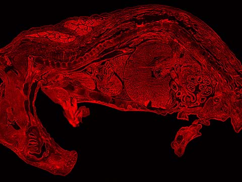
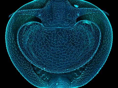
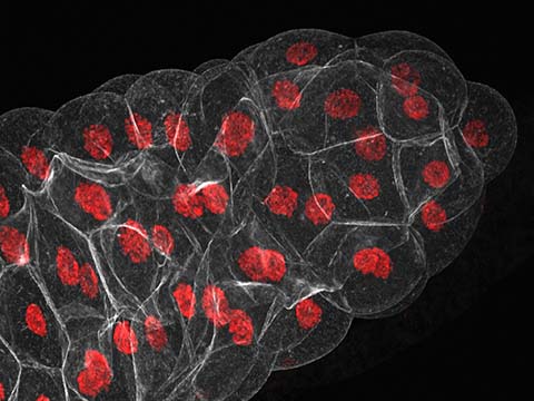
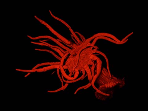
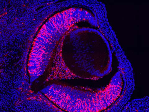
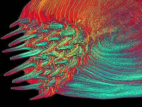
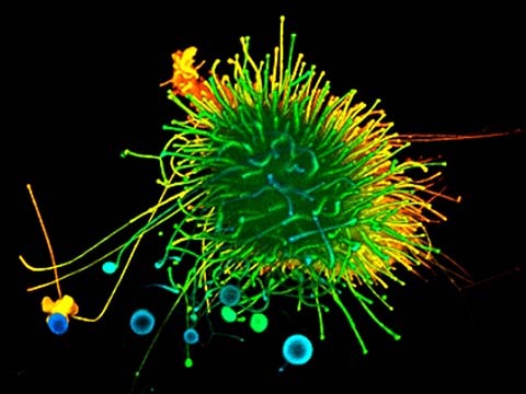
Live zebrafish embryo at 22 hours post-fertilization
Dr. Philipp Keller
- Affiliation
- Howard Hughes Medical Institute (HHMI)
Ashburn, Virginia, USA
- Technique
SiMView Light-Sheet Microscopy
Top 20

Honorable Mentions
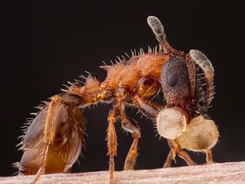
Images of Distinction

Judges
Michael Davidson
Director, Optical and Magneto-Optical Imaging Center at the National High Magnetic Field Laboratory Florida State University
Michael Davidson is the director of the Optical and Magneto-Optical Imaging Center at the National High Magnetic Field Laboratory at Florida State University. Involved with various aspects of microscopy for over 25 years, Davidson’s scientific interests include the packaging of DNA into virus heads, liquid crystallinity in biological systems and the adsorption of small liquid crystal molecules onto surfaces. Davidson has authored many scientific articles on the subject of photomicrography and his photomicrographs have been published in more than a thousand national and international scientific journals, popular magazines and newspapers. In addition, Davidson’s photomicrography has won more than 40 awards in scientific and industrial photography competitions and has been exhibited at over 50 locations nationwide. He is also the expert behind the Nikon Instruments educational Web site MicroscopyU (which can be accessed through the Nikon Instruments Web site at www.nikoninstruments.com) and his own www.molecularexpressions.com.
Laura Helmuth, Ph.D.
Science Editor Slate
Laura Helmuth is Slate’s health and science editor. She was previously the science editor for Smithsonian magazine and an editor for Science magazine’s news department. She has a Ph.D. in cognitive neuroscience from the University of California at Berkeley. She serves on the boards of The Open Notebook and the National Association of Science Writers.
Paul Maddox, Ph.D.
Assistant Professor and William Burwell Harrison Fellow University of North Carolina at Chapel Hill, Department of Biology
Paul S. Maddox, Ph.D. is an Assistant Professor and William Burwell Harrison Fellow, Department of Biology, University of North Carolina at Chapel Hill, North Carolina, USA. Dr. Maddox completed his Ph.D. under the mentorship of renowned cell biology microscopist E. D. Salmon at the University of North Carolina at Chapel Hill. His current research interests include using light microscopy to understand chromosome and microtubule dynamics. Dr. Maddox’s experimental philosophy mandates using the light microscope to “see” results in the most quantitative manner possible. Dr. Maddox has over 19 years of experience with light microscopy and has been an instructor in light microscopy courses around the world including Europe, South America, and at the Marine Biological Labs in Woods Hole, USA. He has published over 60 peer-reviewed papers in his career.
Dave Mosher
Online Director Popular Science
Dave Mosher is the online director of Popular Science, the world’s largest science and technology magazine. He is a journalist with a biology degree whose work has appeared in WIRED, Scientific American, Popular Mechanics, Discover, Space.com, National Geographic News, Discovery.com, and other outlets. In his reporting adventures, Mosher has watched humans and robots launch into space, chronicled crazy home-built contraptions, toured defunct nuclear reactors, and open-sourced his microbiome in the name of science.
Eric Clark
Optical and Magneto-Optical Imaging Center at the National High Magnetic Field Laboratory, Florida State University
Eric Clark has been with Florida State University’s National High Magnetic Field Laboratory (NHMFL) since 1999. Eric serves as webmaster and applications coordinator for the Office of Research and Optical Microscopy division at the NHMFL. Prior to working at FSU, Eric was a surgical nurse and wound care specialist for ten years.