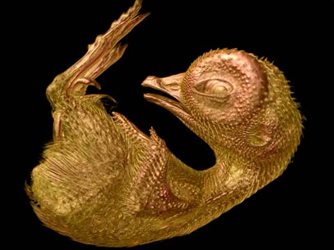
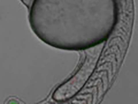
This 3D reconstruction of a quail embryo – comprised of more than 1,000 separate images – shows in startling clarity and detail the anatomy of the specimen. The winning video shows a sequence of “virtual” slices through the whole embryo with 10 days of (in egg) gestation. With this technique, studying the whole anatomy of large specimens like this embryo (23mm long) is possible.
2013 Small World In Motion Competition
1st Place
Quail Embryo at 10 Day Incubation (3D reconstruction)
Dr. Gabriel G. Martins
- Affiliation
- University of Lisbon
Instituto Gulbenkian de Ciencia & CBA/Faculdade de Ciencias Universidad de Lisboa
Lisbon, Portugal
- Technique
Optical tomography, illuminated with a blue LED light (green fluorescence)
- Magnification
- 1x
2nd Place
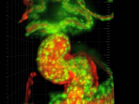
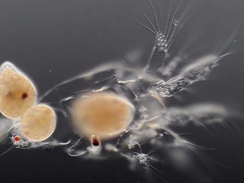
Heart of a two day old zebrafish
Dr. Michael Weber
- Affiliation
- Max Planck Institute of Molecular Cell Biology and Genetics
Dresden, Saxony, Germany
- Technique
Light Sheet Microscopy (Selective Plane Illumination Microscopy)
- Magnification
- 20x
This video shows the beating heart of a two-day old zebrafish embryo. The heart – which is only 250 micrometers or just a bit more than the diameter of a human hair – was reconstructed in 3D after being captured using light sheet fluorescence microscopy in the living zebrafish. In one of its more mesmerizing facets, viewers can watch the movement of blood cells through the heart and its adjacent vessels.
Top 20
Honorable Mentions
Judges
Cara Santa Maria
Science Educator

Cara Santa Maria has dedicated her life to improving science literacy by communicating scientific principles across media platforms. A North Texas native, she currently lives in Los Angeles. Prior to moving to the West coast, Cara taught biology and psychology courses to university undergraduates and high school students in Texas and New York.
Her published research has spanned various topics, including clinical psychological assessment, the neuropsychology of blindness, neuronal cell culture techniques, and computational neurophysiology. Cara previously worked as the senior science correspondent for The Huffington Post, where she wrote, produced, and hosted a weekly video series called “Talk Nerdy To Me.” She also co-stars in “Hacking The Planet” and “The Truth About Twisters” on The Weather Channel.
Cara has appeared on Larry King Live (CNN), Parker/Spitzer (CNN), Geraldo at Large (Fox News), I Kid (TLC), The War Room (Current TV), The Nerdist (BBC America), The Young Turks (Current TV), and Attack of the Show (G4). She co-produced/hosted a pilot for HBO, and she has been a guest on multiple episodes of StarTalk Radio with Dr. Neil deGrasse Tyson, The Joe Rogan Experience, and The Nerdist podcasts.
Michael Davidson
Director, Optical and Magneto-Optical Imaging Center at the National High Magnetic Field Laboratory Florida State University
Michael Davidson is the director of the Optical and Magneto-Optical Imaging Center at the National High Magnetic Field Laboratory at Florida State University. Involved with various aspects of microscopy for over 25 years, Davidson’s scientific interests include the packaging of DNA into virus heads, liquid crystallinity in biological systems and the adsorption of small liquid crystal molecules onto surfaces. Davidson has authored many scientific articles on the subject of photomicrography and his photomicrographs have been published in more than a thousand national and international scientific journals, popular magazines and newspapers. In addition, Davidson’s photomicrography has won more than 40 awards in scientific and industrial photography competitions and has been exhibited at over 50 locations nationwide. He is also the expert behind the Nikon Instruments educational Web site MicroscopyU (which can be accessed through the Nikon Instruments Web site at www.nikoninstruments.com) and his own www.molecularexpressions.com.