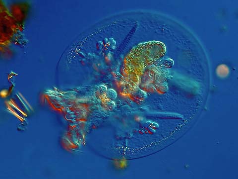
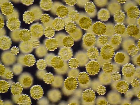
This video is one part of a series Wim van Egmond calls “Swarms,” featuring a colony of golden algae with two flagella swarming toward the light, moving in a mesmerizing pattern reminiscent of constellations. To capture the movement and give depth to the image, he was careful to provide plenty of space for the organisms to move freely. The organisms in the background are a bit out of focus, giving depth more realistic to their natural habitat. “It’s like an abstract painting that moves,” says van Egmond. “That’s what I love about this. Just masses of microbes filling the view, forming patterns as they congregate toward the light.”
2013 Small World In Motion Competition
Honorable Mention
Colonial chrysophytes, Synura uvella
Wim van Egmond
- Affiliation
- Micropolitan Museum
Berkel en Rodenrijs, Zuid-Holland, The Netherlands
- Technique
- Differential Interference Contrast
Honorable Mention
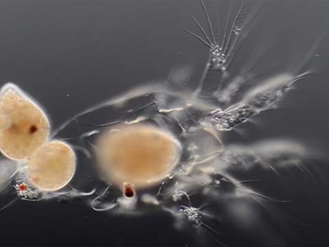
Ophryoglena atra (ciliates) feasting on a copepod larva
Wim van Egmond
- Affiliation
- Micropolitan Museum
Berkel en Rodenrijs, Zuid-Holland, The Netherlands
- Technique
- Differential Interference Contrast
- Magnification
- 160x
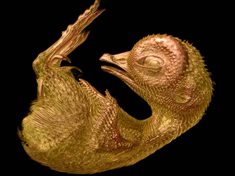
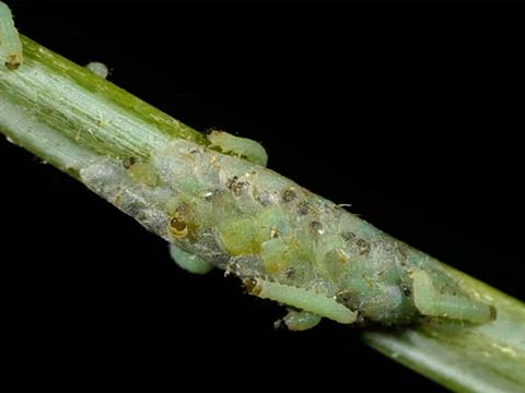
This video plays out more like a microscopic thriller, with two ciliates scavenging inside the body of a dead organism. Like vultures, ciliates detect dying animals on which to feed. In this case the “victim” is the larvae of a copopod – although how they entered the body is a mystery to van Egmond, who was surprised to capture the pair on their “great escape” once they finished feeding.
Capturing such a scene took a combination of patience and luck, as van Egmond carefully laid out his slides, examining them every so often. He first took shots focusing on the ciliates feeding on the organic material. Thirty minutes passed before he returned to examine the slides, and just managed to catch them fleeing the scene. “It’s strange and beautiful, these simple organisms have no eyes, no ears, no organs,” says van Egmond. “But still, they sense one another. One escapes, and the other follows in an instant. It’s fascinating.”
Honorable Mention
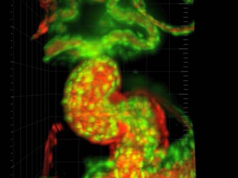
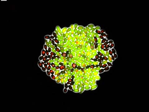
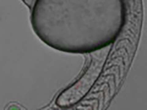
Real-time imaging of the segmentation clock
Dr. Daniele Soroldoni
- Affiliation
- MRC National Institute for Medical Research
London, United Kingdom
- Technique
Multi-dimensional (xyzt) wide-field microscopy
- Magnification
- 10x
For any vertebrate animal, “segmentation,” or the biological division of an animal into repetitive segments such as ribs and vertebrae, is essential to ensure proper physique. Despite its necessity to the vertebrae body plan, segmentation is all too often overlooked for more glamorous processes. Dr. Daniele Soroldoni, this month’s Nikon Small World in Motion highlight, promotes an appreciation for this phenomenon by bringing it into focus beneath the microscope.
Segmentation is notoriously difficult to visualize due to its occurrence during embryo development, so Dr. Soroldoni sought to visualize the patterning sequence at the molecular level. His resulting video beautifully illustrates the pattern formation during segmentation, to show both when and where each vertebrae segment is formed. The clip actually expresses the point in time (denoted by the green wave) in which temporal information is translated into spatial information. Where the green wave stops, the red gene appears in the position where the next segment boundary is formed, ultimately displaying the vertebrates’ growth processes.
Dr. Soroldoni accomplished this capture in his research lab at University College London by delicately embedding fluorescent protein into the chromosome of a zebrafish to create a fusion among the genes of interest. He then tagged the genes in red and green to enable tracking. Using multi-dimensional wide-field microscopy, he combined a large field of view with a highly sensitive and fast EM-CCD camera which enabled him to detect and follow the very faint and transient activity of multiple BAC* transgenes simultaneously.
Ultimately Dr. Soroldoni’s video not only shows a beautiful glimpse into a rarely noticed biological process – but highlights the toolbox he developed for the scientific community, opening the door for continued knowledge and understanding of a complex genetic network.
* A bacterial artificial chromosome (BAC) is an engineered DNA molecule used to clone DNA sequences in bacterial cells. [Source]
Top 20
Honorable Mentions
Judges
Cara Santa Maria
Science Educator

Cara Santa Maria has dedicated her life to improving science literacy by communicating scientific principles across media platforms. A North Texas native, she currently lives in Los Angeles. Prior to moving to the West coast, Cara taught biology and psychology courses to university undergraduates and high school students in Texas and New York.
Her published research has spanned various topics, including clinical psychological assessment, the neuropsychology of blindness, neuronal cell culture techniques, and computational neurophysiology. Cara previously worked as the senior science correspondent for The Huffington Post, where she wrote, produced, and hosted a weekly video series called “Talk Nerdy To Me.” She also co-stars in “Hacking The Planet” and “The Truth About Twisters” on The Weather Channel.
Cara has appeared on Larry King Live (CNN), Parker/Spitzer (CNN), Geraldo at Large (Fox News), I Kid (TLC), The War Room (Current TV), The Nerdist (BBC America), The Young Turks (Current TV), and Attack of the Show (G4). She co-produced/hosted a pilot for HBO, and she has been a guest on multiple episodes of StarTalk Radio with Dr. Neil deGrasse Tyson, The Joe Rogan Experience, and The Nerdist podcasts.
Michael Davidson
Director, Optical and Magneto-Optical Imaging Center at the National High Magnetic Field Laboratory Florida State University
Michael Davidson is the director of the Optical and Magneto-Optical Imaging Center at the National High Magnetic Field Laboratory at Florida State University. Involved with various aspects of microscopy for over 25 years, Davidson’s scientific interests include the packaging of DNA into virus heads, liquid crystallinity in biological systems and the adsorption of small liquid crystal molecules onto surfaces. Davidson has authored many scientific articles on the subject of photomicrography and his photomicrographs have been published in more than a thousand national and international scientific journals, popular magazines and newspapers. In addition, Davidson’s photomicrography has won more than 40 awards in scientific and industrial photography competitions and has been exhibited at over 50 locations nationwide. He is also the expert behind the Nikon Instruments educational Web site MicroscopyU (which can be accessed through the Nikon Instruments Web site at www.nikoninstruments.com) and his own www.molecularexpressions.com.