2013 Small World In Motion Competition

An egg mass of the spruce budworm, Choristoneura occidentalis, hatching over a 2-hour time period. The egg mass is affixed to a needle of a Douglas fir tree

Dr. Ward B. Strong

Affiliation
BC Ministry of Forests, Lands and Natural Resource Operations
Vernon, British Columbia, Canada
Technique
Stereomicroscopy with time-lapse photography
Magnification
15x

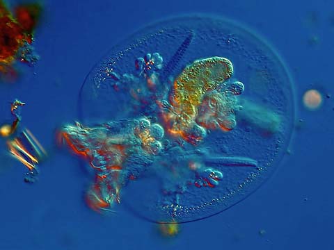
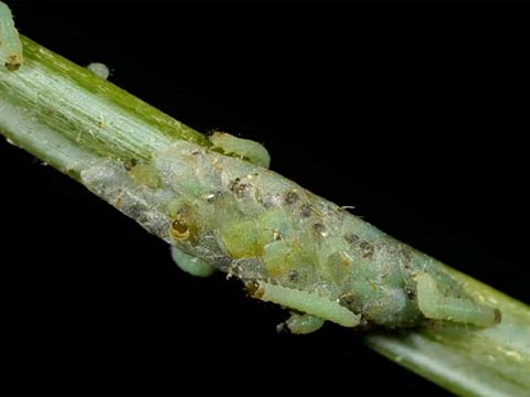
To the naked eye, there’s nothing exciting or even noticeable about the tiny egg mass on a spruce needle. But look a little closer, at just the right time, and you might just see something fascinating.

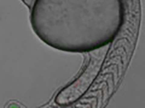
Dr. Ward Strong of Vernon, Canada happened to be in the right place at the right time, looking at a specific spruce needle. The result? An enthralling stop-motion video featuring fifty spruce budworms hatching simultaneously from an egg mass, chewing their way out as individual larvae. Through use of a Greenough Stereo Microscope at 15x magnification paired with a Nikon D90 controlled with a computer to take pictures every five seconds, Dr. Strong was able to capture about 1400 images, which he then assembled into a movie at twenty frames per second. “What I think is amazing is when you’re looking at these by eye, in real time, it happens very slowly, over two hours. It’s an inconspicuous egg mass on a spruce needle; you wouldn’t notice it,” says Dr. Strong. “But speed it up, you see how truly fascinating it is. Simultaneously it froths and bubbles, as each budworm begins squirming and wiggling to escape.”

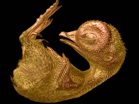
Spruce budworms lay their eggs in summer and hatch in August, producing only a single generation each year. Once they escape the egg mass, each tiny larva burrows into a needle and spends the winter there, safe from natural enemies and the elements. In spring, it resumes feeding on the tree. It’s these feeding habits that caught the attention of Dr. Strong and his research team, who for the past three years have studied the genomics of pest resistance in trees in collaboration with University of British Columbia in Vancouver.

Thanks to increasingly rapid climate change, trees are becoming less and less adapted to the places they live – and therefore more susceptible to pests and insects such as spruce budworms.  While some trees are more resistant to pests than others, Dr. Strong’s team noted that the current breeding system is too slow to keep up with the changing climate.  They set out to assist this process with genomic marker assisted selection – a brand-new mechanism they hope will someday improve breeding in trees that are resistant to pests.  Spruce budworms such as those captured in Dr. Strong’s winning entry have been vital to this study, as researchers observed which trees the budworms prefer to feed upon, and which trees are resistant.  Identifying markers in the tree DNA that correlate with weevil resistance will ultimately speed the process of breeding weevil-resistant trees, thus improving our ability to respond to climate change. As a model system for a new technology, this project also promises applications in breeding for any desirable trait in trees.

According to Dr. Strong, this winning video captures more than just the research behind it.  “As a scientist with a passion for the arts, photography through microscopes has been a very strong theme in my life and career,” says Dr. Strong.  “I’m enthralled with the opportunity provided by this technology, to show the world what is happening under the microscope. There’s an astounding, beautiful, fascinating world under there… if we only take a moment to look a little closer.”

A rotifer (Testudinella)

Ralph Grimm

Location
Jimboomba, Queensland, Australia
Technique
Differential Interference Contrast

When Ralph Grimm sat down at his microscope in early 2013, he expected a leisurely session filming and studying his favorite aquatic creature, the rotifer, having a swim. Instead, he found an animal trying desperately to detach itself from a piece of detritus, struggling against the weight of the debris. Quickly realizing the tiny drama at play, he switched between several magnifications to zoom in and out of the scene, like a filmmaker might, capturing the act until it reached its ultimate conclusion — the rotifer finally gaining “the upper hand” against its unknowing captor and swimming to freedom.

The combination of technique — using two magnifications to capture both the detail of the struggle and the rotifer up close — and artful staging, earned Grimm an honorable mention in the Nikon Small World in Motion competition. It is this type of sharp eye for aesthetics, combined with skilled scientific know-how, that is the hallmark of the Nikon Small World In Motion competition (and its sister competition, Nikon Small World.)

As a teacher of fine arts at a high school in Queensland, Australia, Grimm is not a scientist by trade. However, he has practiced microscopy since childhood — encouraged first by his parents — and used it to satisfy a lifelong love of learning about the visible, and invisible, world around us.

“There are so many things happening under the microscope that are thoroughly ignored by a lot of people,” says Grimm. “They’re scared of the words education and science, thinking it sounds boring or it’s all viruses and bacteria — creatures that are small and dangerous. However, there is so much inherent beauty and mystery - even just plain cuteness. I liked that my video shows my own humorous take on these themes.”

Grimm was also pleased to showcase his favorite microscopy subject, the rotifer. “Rotifers are fascinating. They’re almost completely transparent, so unlike almost any other creature on earth, their inner organs are completely accessible to deeper observation.”

Additionally, the creatures are incredibly good swimmers — able to maneuver in multiple directions and in varying speeds — seemingly without effort. Says Grimm, “From a bio-technology perspective, the study of rotifers often leads me down new lines of thinking that keep me stimulated and always wanting to learn more. On top of that, the biomechanical principals and mechanisms displayed by rotifers could be used for future designs of submersibles and robotic technology.”

Grimm entered the Nikon Small World In Motion Competition, because he wanted the general public to understand that the world under the microscope — the struggles, the joy, the humor, seeing life at work—is a microcosm of the world outside. He hopes to change the perception of science education as something scary or boring, and reveal the beautiful, hidden world waiting to be discovered just beyond the naked eye.

“Education is about more than just earning a degree or getting a job. For me, it’s about adventurous enquiry into the world around us that can lead to great discoveries in the future,” says Grimm.  “There’s a hidden world that deserves to be appreciated and recognized as another level of life. Education gives us the power to uncover it.”

Budding yeast with Gal1 promoter driving unstable GFP and myo1-mcherry during induction. Upon switch from glucose to galactose media, GFP is activated stochastically.

Lu Bai

Affiliation
Penn State University
University Park, Pennsylvania, USA

Anyone who’s enjoyed a glass of wine or fresh-baked bread can appreciate the amazing scientific properties of yeast. Yet for scientists such as Lu Bai, yeast is capable of so much more. Bai works with yeast to study gene expression, thanks to how much one can change its genetic content. In the world of genetic research, yeast allows means of experimentation that is much harder with human cells, fruit flies, and other samples.

Bai is Assistant Professor of Biochemistry and Molecular Biology and Assistant Professor of Physics at Pennsylvania State University. She is also one of the Honorable Mention winners of the Nikon Small World in Motion competition for her captivating video, “Budding Yeast.” In her video, we see how this amazing species shows genes expressing themselves in reproducing cells.

Aesthetically, the video is mesmerizing. In it, we see the yeast doubling and growing in size. It becomes hypnotic to watch as it takes on a new form with every new addition. But take a closer look, and like everything in the Nikon Small World universe, there is more than meets the eye.

Though the video is less than 30 seconds in length, what we’re actually viewing is a movie that took place over the course of 10 hours. Every time we see the yeast double (which happens every second of the video or so), we’re actually seeing is about 1.5 hours of real-time work. There is more to this technique than just speeding up the video, however. It’s hard to keep the environment for yeast to be “happy,” as Bai puts it. One has to maintain the environment for growth, while keeping the focus constant and the sample stage stable so the video comes out clearly.

Even more important is the green fluorescence we see in Bai’s video, highlighting the genes she is interested in studying. “All the cells you see here are from the same family: they originate from one ‘mother cell’ and grow from there, so genetically they are all the same,” Bai says. “At the beginning of the movie, we changed the yeast growth conditions, and the cells respond by ‘turn-on’ some genes, and one of them has green fluorescence. In other words, the green color in the cells represent their response to changing environment. If you look carefully, you will notice that different cells respond at very different time, some after two hours, and some after seven hours. In other words, the cells have ‘individuality’ and make different ‘decisions.’ This is the basis of both my research and fascination!”

Bai’s research is important, because in the human body, the same type of cells may also have different gene expressions, and it may sometimes cause disease - like cancer - for example. Understanding how and why individuality occurs, then, is the key to unlocking how to prevent them from occurring in the first place, if needed. To do this, her work involves characterizing the differences among cells, understanding if the individuality that occurs is good or bad, and trying to work out how the cells control these differences. Bai’s research is part of a larger body of work being done that hopes to uncover why these phenomenon take place. It can only be done studying cells one-by-one like we see here.

In Bai’s video, we see the work to solve one of modern science’s current mysteries. Her contribution to Nikon Small World in Motion showcases the best of the competition - a passion for science, shown through incredible research, and in a way that is aesthetically pleasing to the eye and that the general public can start to understand.

Bai says that while the scientific community started by studying yeast, they are working toward addressing similar questions in higher species. Simply put she says, “We are trying to understand the mechanism so that one day we can learn to control it.”