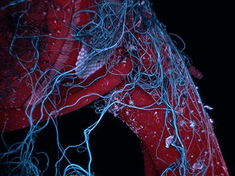
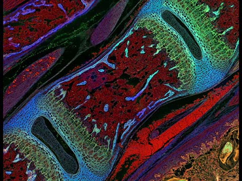
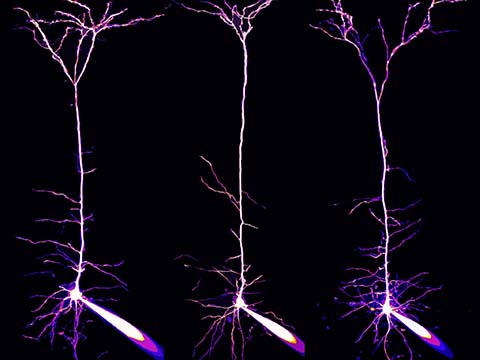
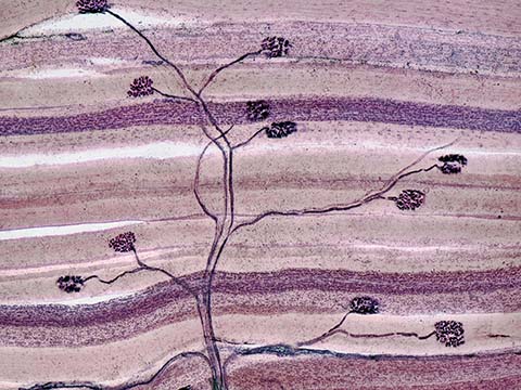
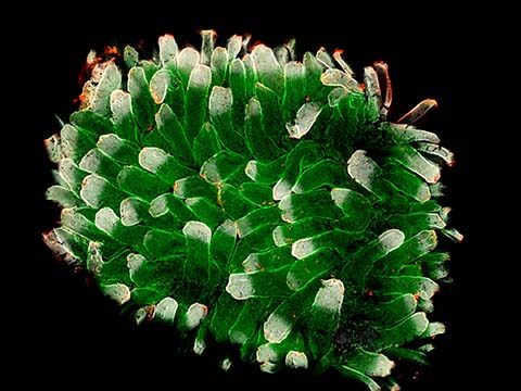
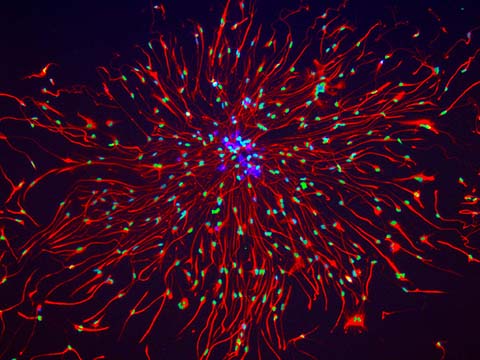
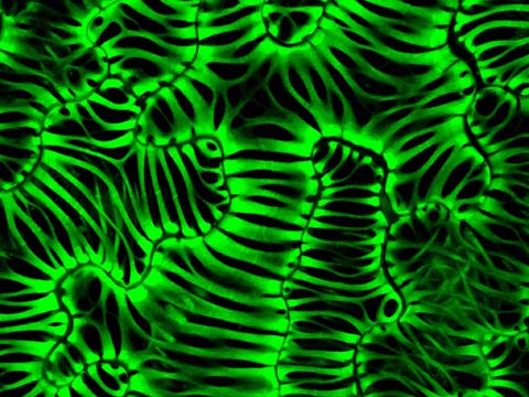
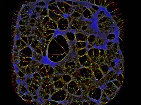
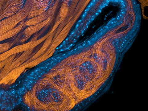
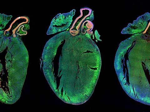
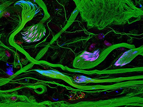
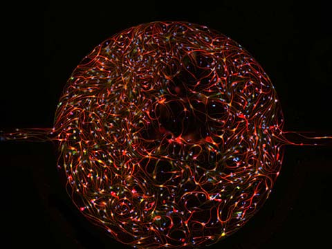
This image shows sections of rat hearts. The muscle cells of the heart, called cardiomyocytes, are stained in green, nucleus of cells are counterstained in blue and proliferating cells are labeled in green.
2013 Photomicrography Competition
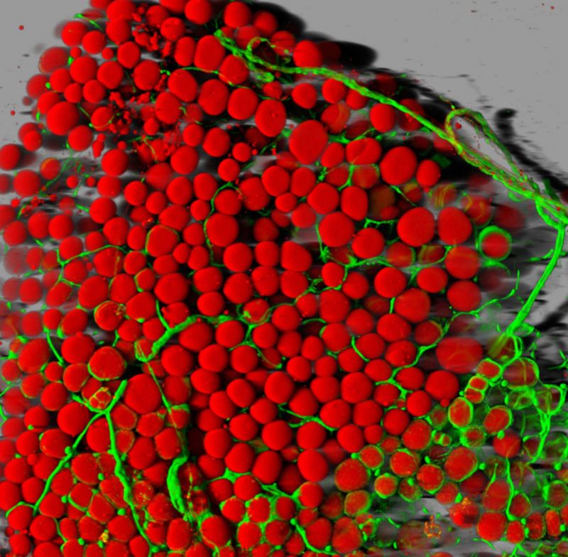
Image of Distinction
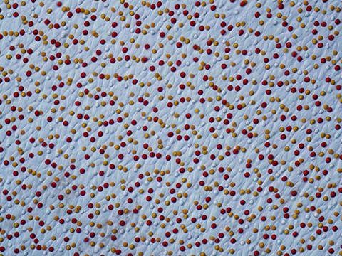
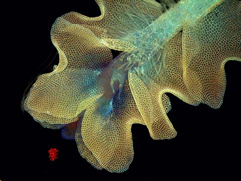
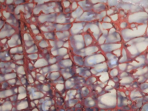
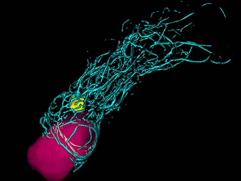
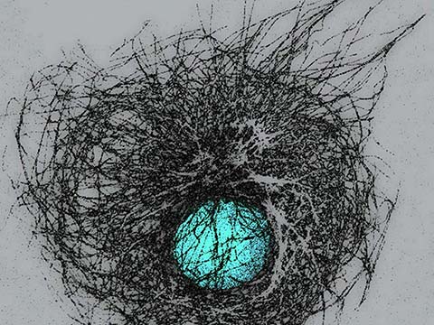
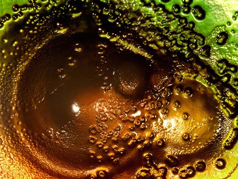
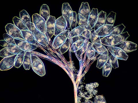
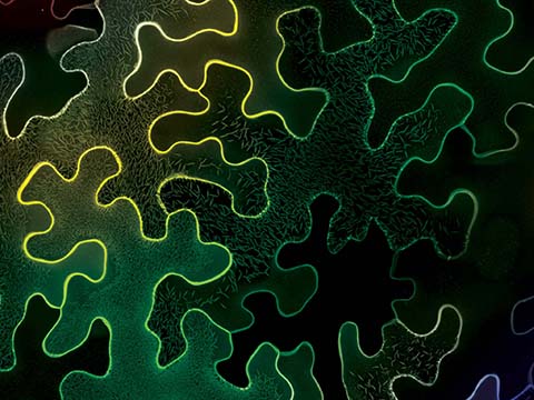
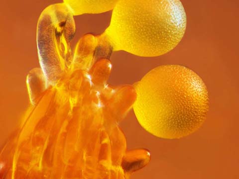
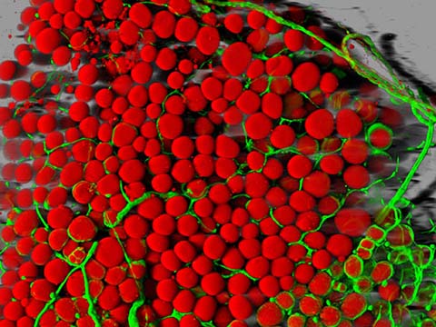
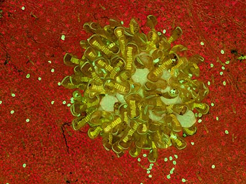
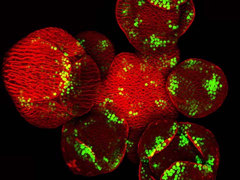
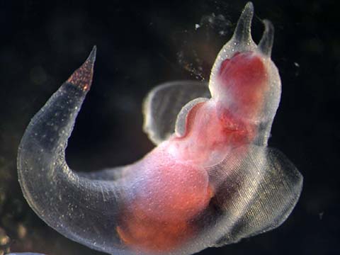
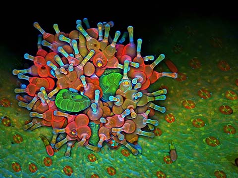
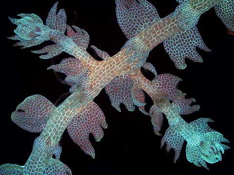
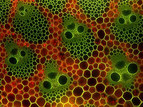
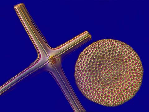
Mouse adipose (fat) tissue
Dr. Daniela Malide
- Affiliation
- National Institutes of Health (NIH)
National Heart, Lung and Blood Institute, Light Microscopy Core Facility
Bethesda, Maryland, USA
- Magnification
- 25x
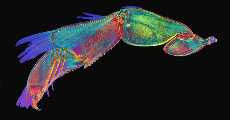
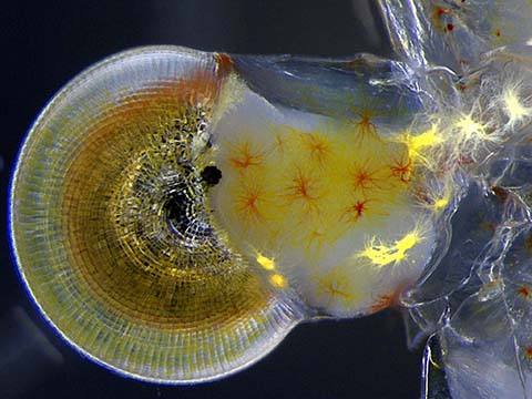
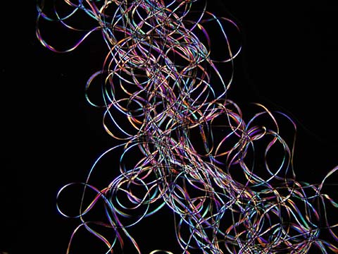
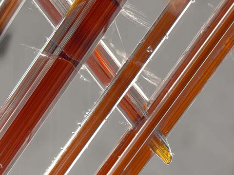
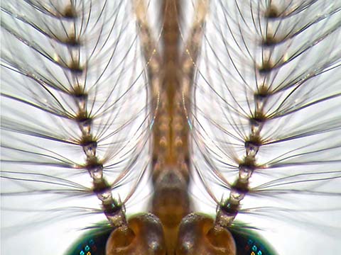
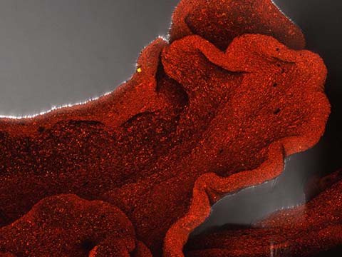
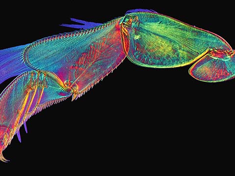
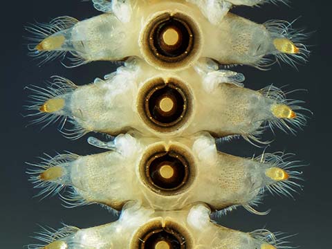
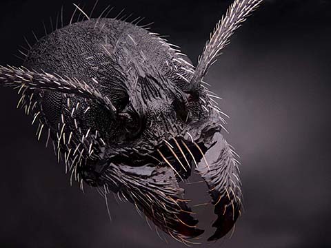
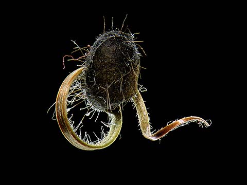
Image of Distinction
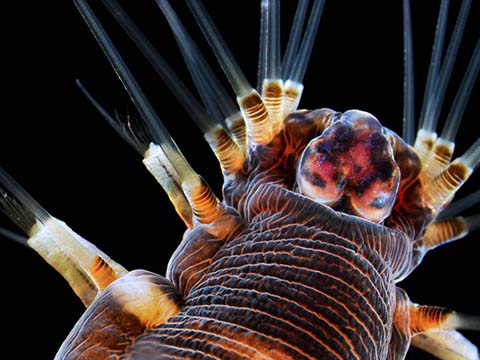
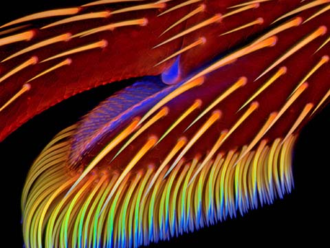
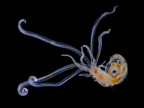
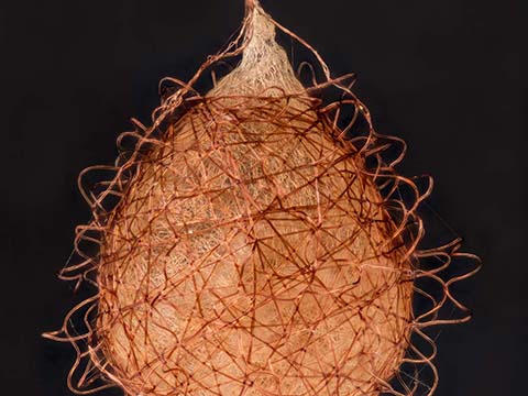
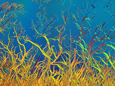
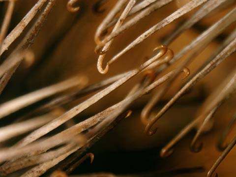
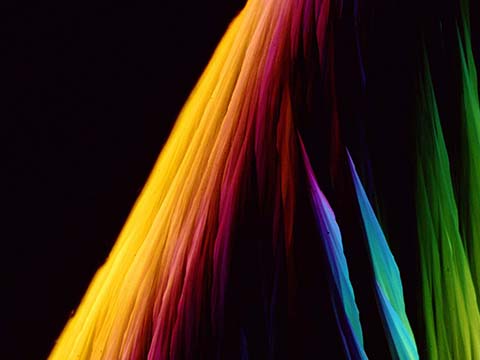
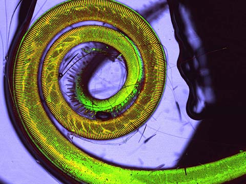
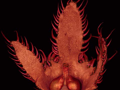
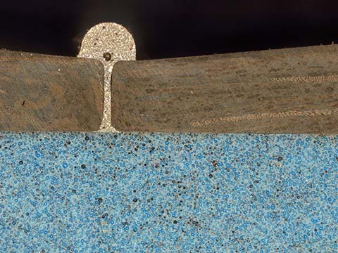
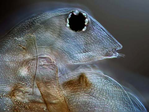
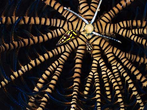
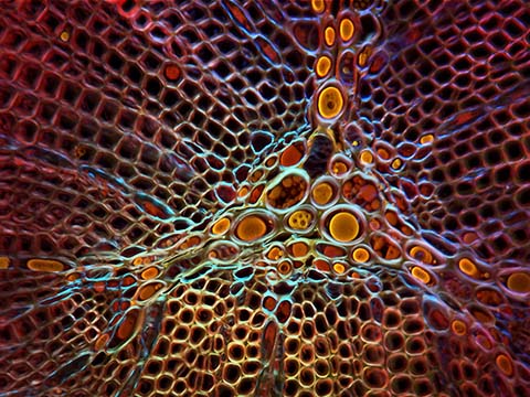
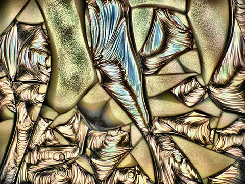
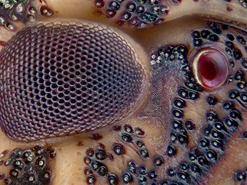
Gyrinus sp. (whirligig beetle) swimming leg
David Linstead
- Location
- Bromley, Kent, United Kingdom
- Technique
- Differential Interference Contrast
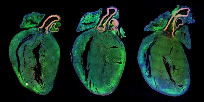
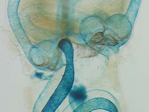
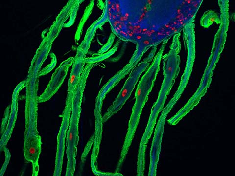
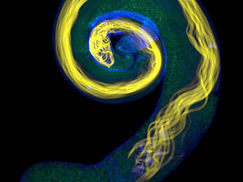
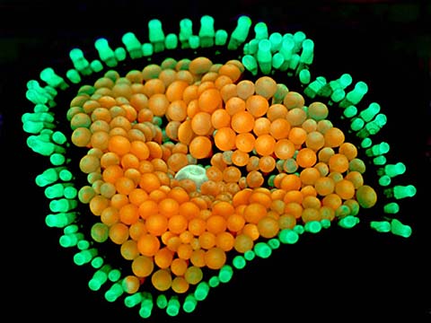
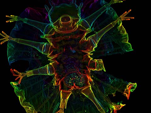
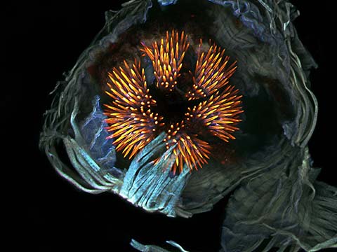
Image of Distinction
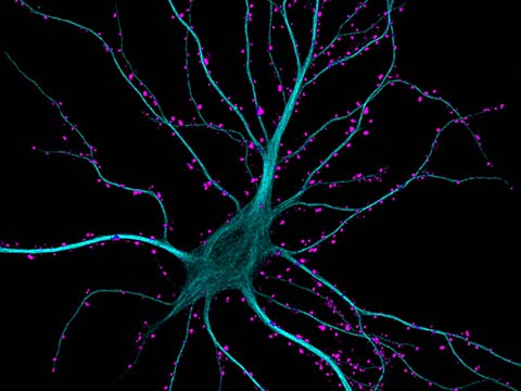
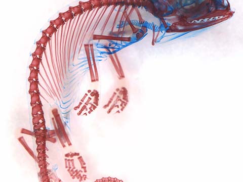
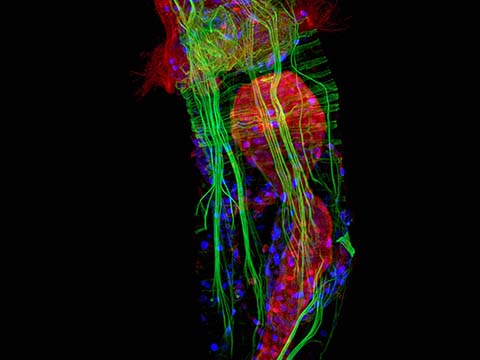
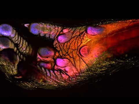
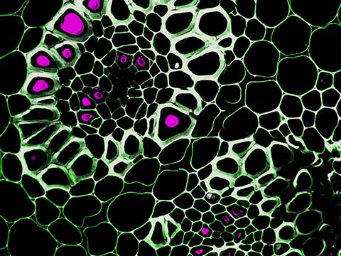
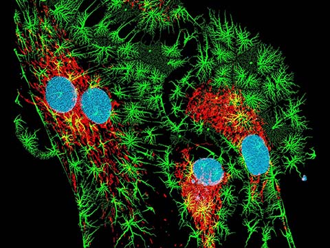
Rat heart sections
Dr. Miguel Mano
- Affiliation
- International Centre for Genetic Engineering and Biotechnology
Department of High-Throughput Screening
Trieste, Italy
- Technique
- Fluorescence
- Magnification
- 4x
Top 20
Honorable Mentions
Images of Distinction
Judges
Dr. Joan V. Ruderman
President and Director Marine Biological Laboratory
Joan V. Ruderman is the 14th director of the Marine Biological Laboratory and the Marion V. Nelson Professor of Cell Biology at Harvard Medical School.
Ruderman has served on the Board of Trustees of the Marine Biological Laboratory (MBL) since 1986. Since 2008, she has been the Speaker of the MBL Corporation and a member of the MBL’s executive committee. Her long affiliation with the MBL began in 1974 when, the summer after receiving her Ph.D., she took the MBL’s Embryology course. She subsequently taught in that course for several years and then, in 1980, returned as a visiting researcher, working at the MBL for most summers thereafter until 2000.
Ruderman is best known for her work on the molecular mechanisms that regulate cell division. In the 1980s and 1990s, she conducted pioneering research on cyclins, proteins that drive cells through the cell division cycle. More recently, Ruderman has investigated environmental contaminants that can mimic estrogen, chemicals that may increase the risk of developing breast cancer and other hormone-dependent cancers.
Ruderman’s scientific contributions have been recognized by her election to the U.S. National Academy of Sciences, the American Academy of Arts and Sciences, and the American Society for Microbiology, as well as by the New York University/Dart Award in Biotechnology. Among her national leadership roles, she has served on the Scientific Advisory Board of Whitehead Institute for Biomedical Research at M.I.T. and the Medical Advisory Board of Howard Hughes Medical Institute. She is an incoming member of the Scientific Advisory Board of the Ellison Medical Foundation (2013-).
Cara Santa Maria
Science Educator

Cara Santa Maria has dedicated her life to improving science literacy by communicating scientific principles across media platforms. A North Texas native, she currently lives in Los Angeles. Prior to moving to the West coast, Cara taught biology and psychology courses to university undergraduates and high school students in Texas and New York.
Her published research has spanned various topics, including clinical psychological assessment, the neuropsychology of blindness, neuronal cell culture techniques, and computational neurophysiology. Cara previously worked as the senior science correspondent for The Huffington Post, where she wrote, produced, and hosted a weekly video series called “Talk Nerdy To Me.” She also co-stars in “Hacking The Planet” and “The Truth About Twisters” on The Weather Channel.
Cara has appeared on Larry King Live (CNN), Parker/Spitzer (CNN), Geraldo at Large (Fox News), I Kid (TLC), The War Room (Current TV), The Nerdist (BBC America), The Young Turks (Current TV), and Attack of the Show (G4). She co-produced/hosted a pilot for HBO, and she has been a guest on multiple episodes of StarTalk Radio with Dr. Neil deGrasse Tyson, The Joe Rogan Experience, and The Nerdist podcasts.
Alan Taylor
Senior Editor The Atlantic
Alan Taylor runs the news photo blog “In Focus” for The Atlantic magazine.
In 2008, Taylor created the successful photo blog “The Big Picture” for The Boston Globe, which he then ran for two years, picking up two Webby Awards and a SXSW nomination for “Best Blog” along the way.
Taylor has played an enormous role in kick-starting the large-photo format blogging trend among news organizations. Formerly a web developer, Taylor combined his love of storytelling and photography with the web skills he’d developed to create a new platform for visual storytelling online.
He covers a wide range of subjects, from breaking news and historical topics to culture.
Dr. Ronald Vale
The University of California – San Francisco
Ronald D. Vale is Professor of the Dept. of Cellular and Molecular Pharmacology at the University of California, San Francisco and an Investigator in the Howard Hughes Medical Institute.
Vale received B.A. degrees in Biology and Chemistry from the University of California, Santa Barbara in 1980. Dr. Vale received his Ph.D. in Neuroscience from Stanford University in 1985 where he trained with Dr. Eric Shooter. He was a Staff Fellow with the N.I.H. stationed at the Marine Biological Laboratory in 1985-6 (with Tom Reese) and began his faculty appointment at UCSF in 1987. During his time at UCSF, he has served as the Director of the Cell Biology Program (4 years) and Vice-Chair (10 years) and Chair of the Dept. of Cellular and Molecular Pharmacology (5 years). He also holds an Adjunct Senior Scientist appointment with the Marine Biological Laboratory at Woods Hole.
Vale founded and produces iBioSeminars, lectures by leading biologists which are made freely available on the web. Vale co-directed the 7 week Physiology Course at the Marine Biological Laboratory, transforming it to a venue of interdisciplinary research in physics and biology (2004-2008). Vale is active in helping young scientists in India by starting the very popular Young Investigator Meetings as well as an interactive web site (IndiaBioscience.org) for India biologists to obtain information on jobs/grants/collaborations. He serves on HHMI’s Education Advisory Committee and on the Advisory Committee for the Indian Institute of Science and Education in Pune.
Dr. Vale’s awards include the Pfizer Award in Enzyme Chemistry (1991), the Young Investigator Award from the Biophysical Society (1993), and the Wiley Prize in Biomedical Sciences (2012). He was elected to the National Academy of Sciences in 2001 and to the American Academy of Sciences in 2002. He has been the Keith Porter Lecturer (ASCB), the Sunney Chan Lecturer (UCB), Daniel Mazia Lecturer (Stanford), the Lamport Lecturer (U. Washington), the Naidorf Memorial Lecturer (Columbia University), the Davies Lecturer (U. Penn), the Baker Memorial Lecturer (U. Michigan), the UCSF Faculty Research Lecturer, and the Keith Porter Friday Night Lecturer (Marine Biological Laboratory).
Michael Davidson
Director, Optical and Magneto-Optical Imaging Center at the National High Magnetic Field Laboratory Florida State University
Michael Davidson is the director of the Optical and Magneto-Optical Imaging Center at the National High Magnetic Field Laboratory at Florida State University. Involved with various aspects of microscopy for over 25 years, Davidson’s scientific interests include the packaging of DNA into virus heads, liquid crystallinity in biological systems and the adsorption of small liquid crystal molecules onto surfaces. Davidson has authored many scientific articles on the subject of photomicrography and his photomicrographs have been published in more than a thousand national and international scientific journals, popular magazines and newspapers. In addition, Davidson’s photomicrography has won more than 40 awards in scientific and industrial photography competitions and has been exhibited at over 50 locations nationwide. He is also the expert behind the Nikon Instruments educational Web site MicroscopyU (which can be accessed through the Nikon Instruments Web site at www.nikoninstruments.com) and his own www.molecularexpressions.com.