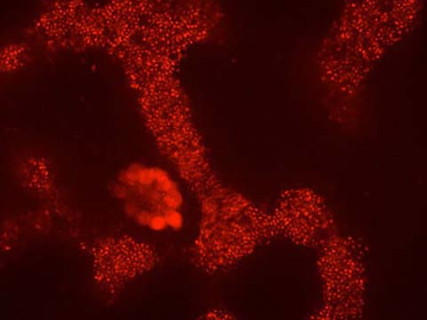
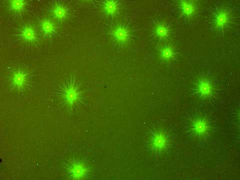
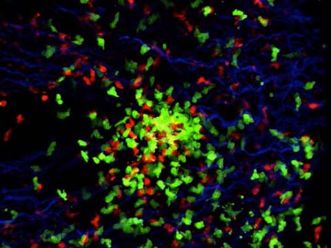
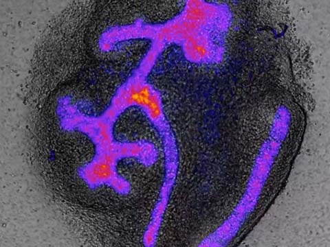
This movie shows the growth, interaction, and movement of microtubule asters (in green) in Xenopus (frog) egg cytoplasm, following exit from metaphase (cell division). Asters grown in a thin layer of cytoplasm between two glass coverslips recapitulates the behavior of asters in early dividing live embryos during anaphase/telophase/cytokinesis. Where asters meet, cytokinesis proteins such as the chromosomal passenger complex (visualized using a fluorescently labeled antibody against a CPC component, shown in red) are recruited. This establishes a boundary between the two asters, and marks the position of the putative cleavage furrow.
2012 Small World In Motion Competition
Honorable Mention
Time lapse movie of microtubule asters growing in a thin layer of interphase Xenopus (frog) egg extract
Phuong Anh Nguyen
- Affiliation
- Harvard Medical School
Mitchison Lab
Department of Systems Biology
Boston, Massachusetts, USA
- Technique
- Widefield fluorescence microscopy
- Magnification
- 10x
Honorable Mention
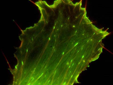
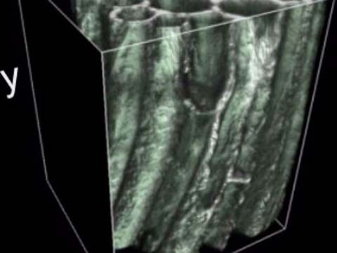
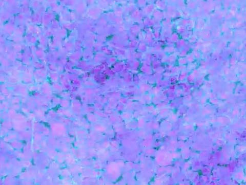
CAR fish fibroblast transfected with mCherry-Actin and GFP-Vasp
Dr. Maria Nemethova
- Affiliation
- IMBA - Institute of Molecular Biotechnology GmbH
Vienna, Austria
- Technique
- Epifluorescence
- Magnification
- 100x
This video illustrates the polymerization of actin, a protein which drives cell movements. Actin acts like the “scaffold” of a cell, so understanding how it forms is important is to basic cellular research and can help answer further questions on how cells form.
Honorable Mention
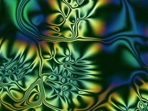
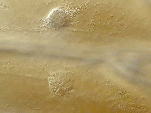
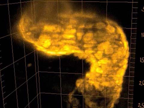
Movement of organelles in plant cells (onion bulb scale epidermis)
Dr. Heiti Paves
- Affiliation
- Tallinn University of Technology
Tallinn, Estonia
- Technique
- Differential Interference Contrast
- Magnification
- 20x
Top 20
Honorable Mentions