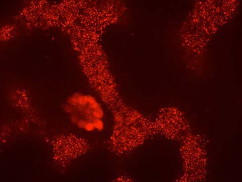
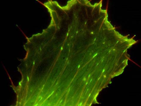
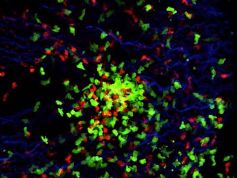
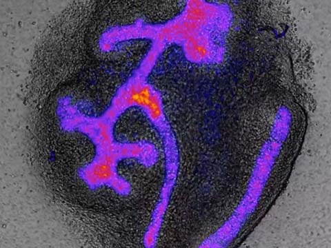
This movie shows the growth, interaction, and movement of microtubule asters (in green) in Xenopus (frog) egg cytoplasm, following exit from metaphase (cell division). Asters grown in a thin layer of cytoplasm between two glass coverslips recapitulates the behavior of asters in early dividing live embryos during anaphase/telophase/cytokinesis. Where asters meet, cytokinesis proteins such as the chromosomal passenger complex (visualized using a fluorescently labeled antibody against a CPC component, shown in red) are recruited. This establishes a boundary between the two asters, and marks the position of the putative cleavage furrow.
2012 Small World In Motion Competition
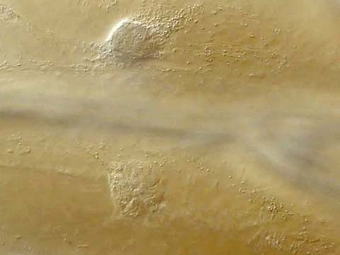
Honorable Mention
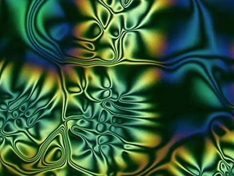
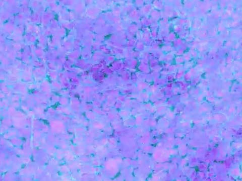
Movement of organelles in plant cells (onion bulb scale epidermis)
Dr. Heiti Paves
- Affiliation
- Tallinn University of Technology
Tallinn, Estonia
- Technique
- Differential Interference Contrast
- Magnification
- 20x
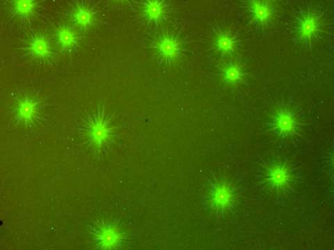
Honorable Mention
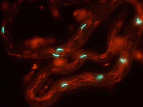
Time lapse movie of microtubule asters growing in a thin layer of interphase Xenopus (frog) egg extract
Phuong Anh Nguyen
- Affiliation
- Harvard Medical School
Mitchison Lab
Department of Systems Biology
Boston, Massachusetts, USA
- Technique
- Widefield fluorescence microscopy
- Magnification
- 10x
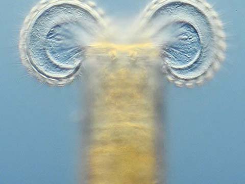
Honorable Mention
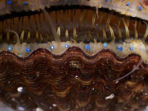
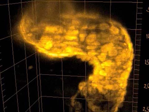
Limnias melicerta (a rotifer)
Wim van Egmond
- Affiliation
- Micropolitan Museum
Berkel en Rodenrijs, Zuid-Holland, The Netherlands
- Technique
- Differential Interference Contrast
- Magnification
- 200x
This microanimal lives in a self-built tube attached to waterplants. We see the rotifer using fast moving cilia to create a vortex. This enables it to sweep in food particles like algae. Inside the organism we can also see jaw-like structures that grind the food.
Top 20
Honorable Mentions