What is the subject matter of your winning entry?
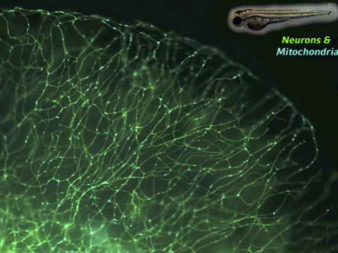
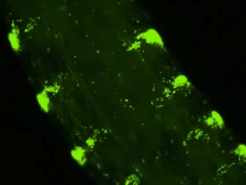
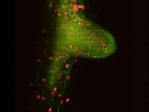
The time-lapse video shows the movement of mitochondria in sensory neurons in the tail of a two-day-old transgenic zebrafish larvae. Two transgenes have been introduced to label the membranes of the neurons with memYFP and the mitochondria with mitoCFP.
What do you think makes this particular movie important/significant or interesting?
It is to our knowledge the first example of live imaging of mitochondrial transport in nerve cells in an intact, unmodified vertebrate animal.
Were there any imaging challenges you faced?
The main problem is the quite rapid movement of the mitochondria, while the fluorescence is rather dim. This not only excludes long exposure times, but also multidimensional confocal imaging. Therefore the imaging was done on a widefield microscope with a very sensitive camera with short exposure times. To avoid a blurry image, this requires mounting the animal in a way that most of the nerve cells are in the same optical plain, which is difficult for an animal, which is only about 1 mm in length.
How does your entry relate to your work?
Our lab studies molecular and cellular pathologies in cell and animal models of Alzheimer’s disease and Frontotemporal Dementia. In both disease alterations of the transport of cellular components are discussed. These transport impairments would cause problems especially to nerve cells, since they are the longest cells in the human body. In particular, the impaired transport of mitochondria could harm the cells, since this organelle is responsible for the generation of energy, leading to energy deficiencies in distant parts of the nerve cells. We cross the fish with the fluorescent mitochondria to our fish models of Alzheimer’s disease and FTD to study transport impairments directly in living animal models of these diseases.

 Share
Share Tweet
Tweet Pin-It