2011 Small World In Motion Competition

Actin dynamics in phagocytosis of budded yeast (Image size: 33 microns x 26 microns)

Dr. Margaret Clarke

Affiliation
Oklahoma Medical Research Foundation
Program in Genetic Models of Disease
Oklahoma City, Oklahoma, USA
Technique
Confocal

Waterflea Daphnia playing with volvox

Dr. Ralf Wagner

Location
Düsseldorf, Germany
Technique
Darkfield
Magnification
50x

What is the subject matter of your winning entry?

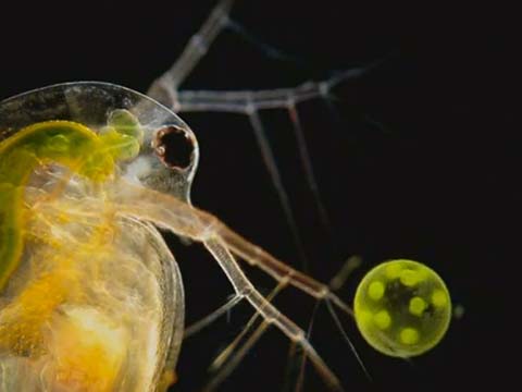
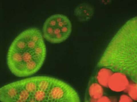
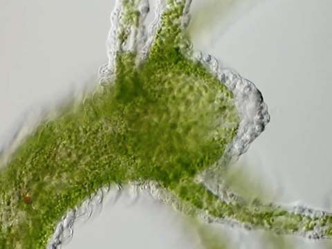
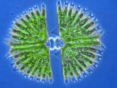
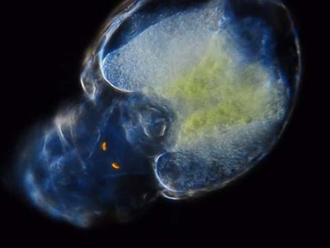
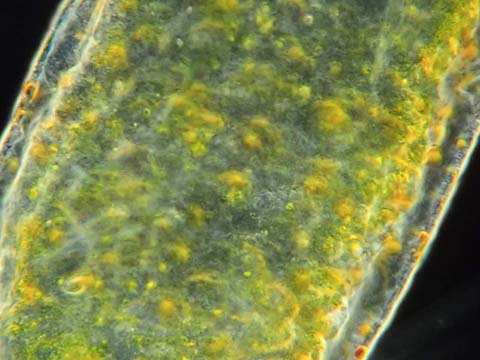
The video shows a daphnia together with a volvox. The volvox is turning and moving along under the slide and at two moments the daphnia is moving its complex-eye towards the direction of the volvox and you get the impression, that the daphnia really is looking at the volvox.

Why did you choose to submit this particular movie?

I thought it was an extraordinary situation, the daphnia looking at a Volvox. The sample for this slide came from my garden pond when there was a bloom of Volvox in it in spring 2011. I decided to play around a little with these Volvox balls and fortunally could capture the situation shown in the movie.

Were there any imaging challenges you faced?

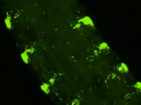
I concentrated the Volvox species with a plankton sieve and took the sample from that concentrate in order to be sure that enough Volvox were in the sample. At the same time of course, the Daphnia were concentrated, too.

An African green monkey kidney cell (COS-7) transfected with mEmerald/cSrc showing membrane ruffles

Dr. Liang Gao

Affiliation
Howard Hughes Medical Institute (HHMI)
Janelia Farm Research Campus
Ashburn, Virginia, USA
Technique
Two Photon Bessel Beam Plane Illumination Microscopy
Magnification
56x

Q&A WITH THE WINNER

What is the subject matter of your winning entry?
Retrograde flow of membrane ruffles formed at the leading edge of an African green monkey kidney cell as well as the macropinocytosis process showing the intracellular vacuoles formation and dynamics.

Why did you choose to submit this particular movie?
This is a good looking cell showing active dynamics.

Can you elaborate on the imaging technique you used?
It’s an example of the imaging quality of the Bessel beam plane Illumination microscopy. A thin excitation light sheet created by a scanning Bessel beam allows fast imaging with excellent three dimensional spatial resolution.