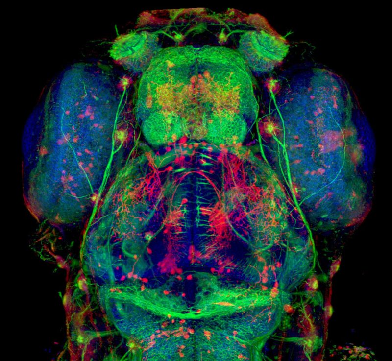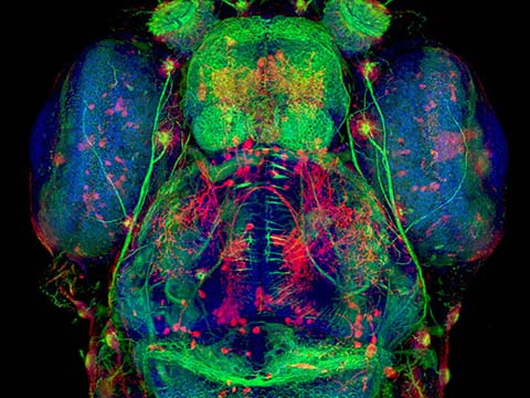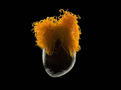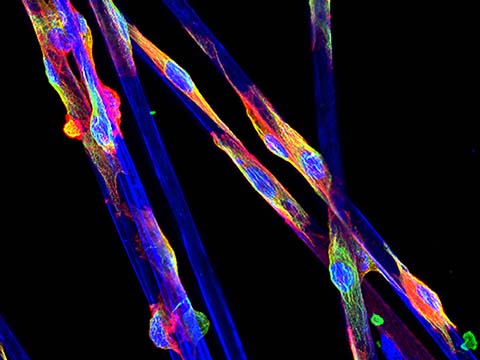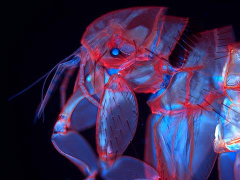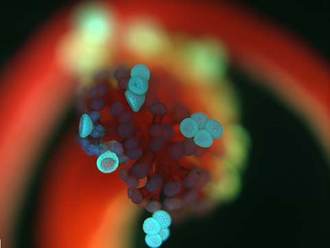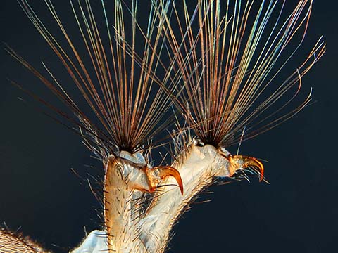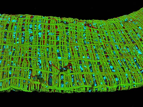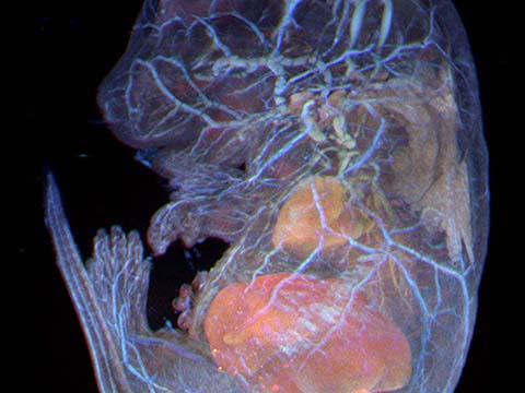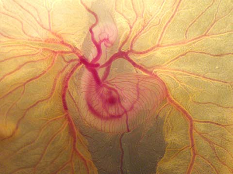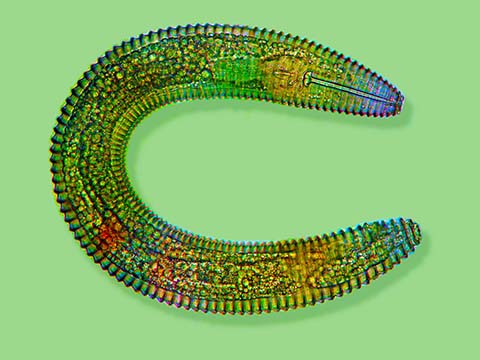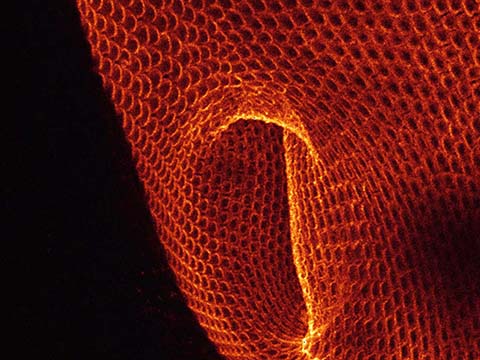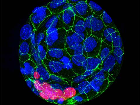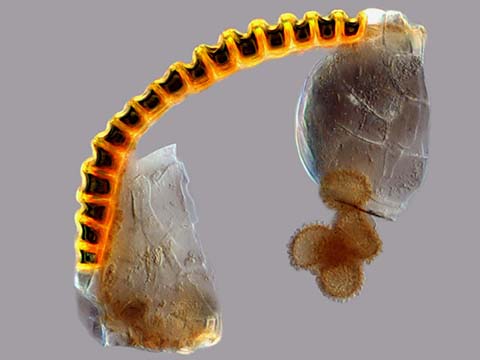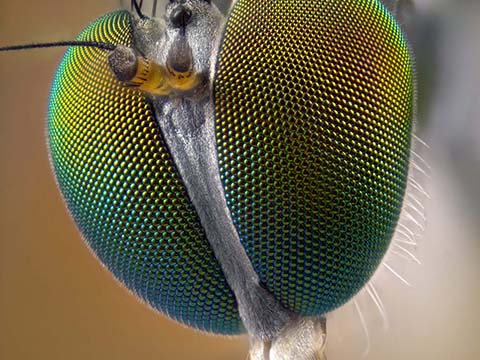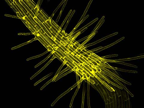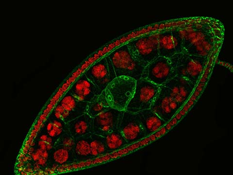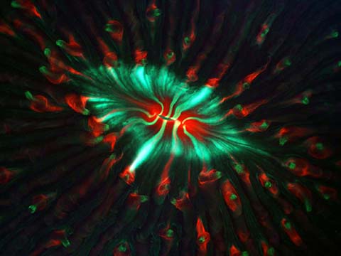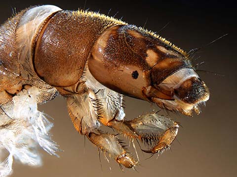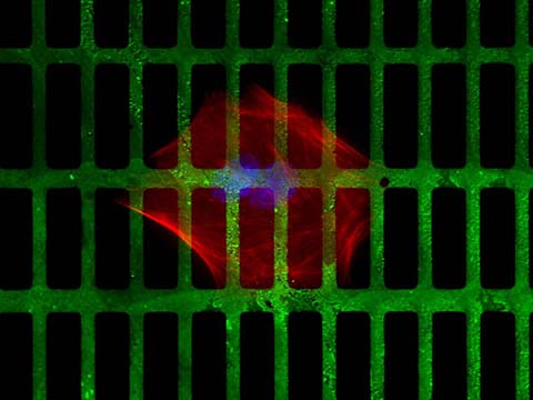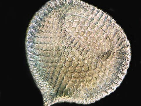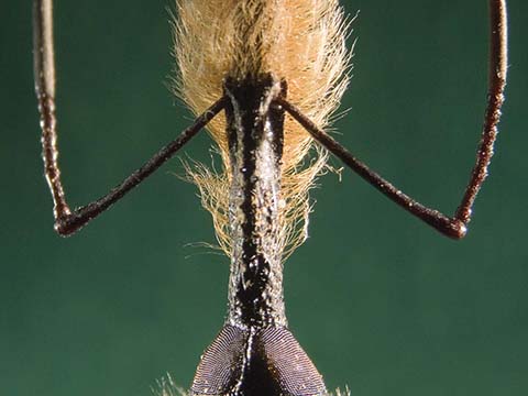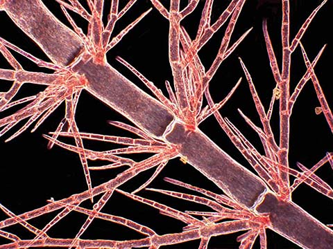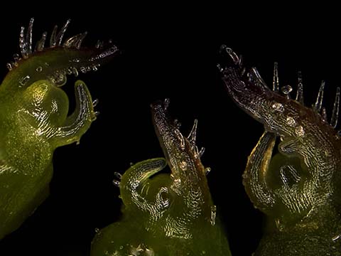
Malaria’s impact worldwide is still an issue, particularly in developing countries. Research is ongoing to study the carriers of malaria, mosquitoes, and how they carry and transmit the disease and other pathogens. That’s why the 2010 winning image by Jonas King is so important to the life science community.
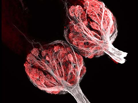
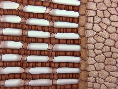
Anopheles gambiae (mosquito heart) was captured at 100x magnification. Jonas works out of Vanderbilt University’s Hillyer Lab, which studies the interactions between mosquitoes and their pathogens, along with salivary components and how they interact with the vertebrate host’s immune response.
The image details the structural organization of the mosquito heart and provides insight into how mosquitoes move blood to all regions of their bodies. Jonas notes, “Mosquitoes remain one of the greatest scourges of mankind. Malaria infects hundreds of millions of people annually and is believed to have a major impact on the economies of endemic regions.”