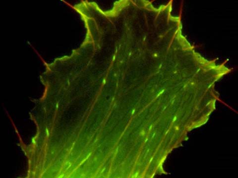
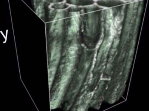
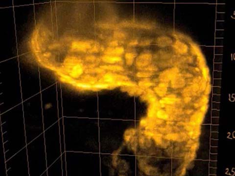
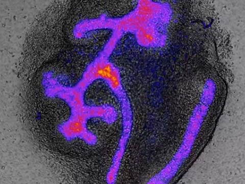
Illustrating the development of live kidney cells, specifically a metanephric kidney that has been cultured in vitro and imaged over 4 days. The fluorescence originates from a conditional YFP reporter, which is only activated in cells expressing Pax8. The fluorescent cells belong to the ureteric bud (the tree), the induced nephron progenitors cell (cells around the tree tips) and nephrons that are forming (the shapes forming within the tree). The YFP signal is viewed using a heat-map that has been overlaid onto the bright-field channel.
2012 Small World In Motion Competition
3rd Place
Complexity of ureteric bud branching and nephron formation
Dr. Nils Lindström
- Affiliation
- University of Edinburgh
The Roslin Institute
Edinburgh, Midlothian, United Kingdom
- Technique
- Time-lapse, inverted fluorescent microscope, organ culture, transgenic mouse reporter lines
2nd Place
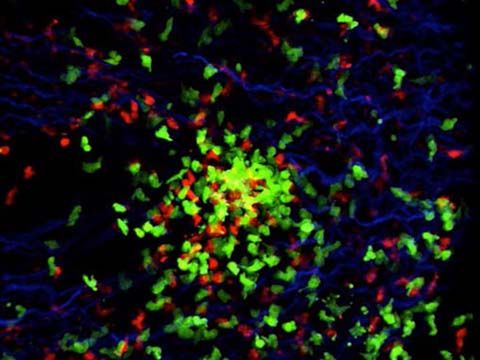
Sperm from two males competing within reproductive tract of a female fruit fly (Drosophila melanogaster)
Dr. Stefan Lüpold
- Affiliation
- Syracuse University
Department of Biology
Syracuse, New York, USA
- Technique
- Fluorescence
- Magnification
- 400x
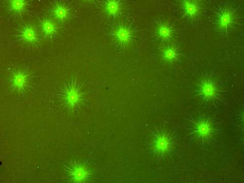
Sperm of two different males (green and red) competing within the female reproductive tract of the fruit fly Drosophila melanogaster. Competition between sperm is a widespread phenomenon throughout the animal kingdom and a powerful evolutionary force driving species diversity. However, it has been nearly impossible to study the fundamental biological processes associated with such sperm competition, occurring whenever sperm from different males mix inside of females. The very recent development of genetically modified fruit flies that produce sperm with either green- or red-fluorescent heads (as seen in the movie) is now allowing us to answer important biological questions.
Honorable Mention
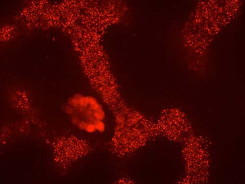
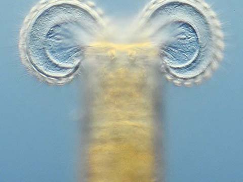
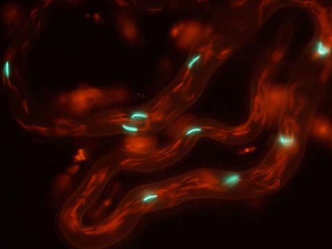
Tetrahymena sp. (ciliate) feeding on a biofilm composed of the bacterium Serratia plymuthica, which is expressing a red fluorescent protein
Andrew Dopheide
- Affiliation
- University of Auckland
School of Biological Sciences
Auckland, New Zealand
- Technique
- Confocal
- Magnification
- 400x
This video shows Tetrahymena sp. (common freshwater protozoa) cells feeding on a biofilm composed of freshwater bacterium Serratia plymuthica. The bacterial cells and biofilm structures are visible due to expression by the bacteria of a red fluorescent protein. Tetrahymena sp. cells are visible as rapidly-moving clusters of intracellular feeding vacuoles packed with red-fluorescing bacterial cells. The focal plane of the video moves down with each successive frame, revealing the effect of Tetrahymena sp. feeding activity on bacterial biofilm morphology as ciliate-sized channels and holes throughout the biofilm.
Top 20
Honorable Mentions